 Back
The Integumentary System: Structure, Function, and Clinical Aspects
Study Guide - Smart Notes
 Tailored notes based on your materials, expanded with key definitions, examples, and context.
The Dermis
Structure and Function of the Dermis
The dermis is the layer of skin beneath the epidermis, providing structural support, nourishment, and housing various accessory structures. It is composed of connective tissue and contains blood vessels, nerves, and glands.
Function: The dermis supports the epidermis, supplies nutrients, and aids in thermoregulation.
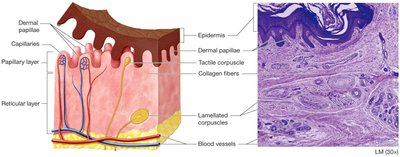
Layers: The dermis is divided into two main layers: the papillary layer (superficial) and the reticular layer (deep).
Papillary Layer
The papillary layer is the uppermost region of the dermis, characterized by thin, loose connective tissue and finger-like projections called dermal papillae.
Dermal papillae: Increase surface area for exchange of gases, nutrients, and waste between dermis and epidermis.
Capillaries: Supply blood to the epidermis.
Tactile corpuscles: Specialized for touch sensation.
Reticular Layer
The reticular layer is the deeper, thicker portion of the dermis, composed of dense irregular connective tissue.
Collagen fibers: Provide strength and resilience.
Lamellated corpuscles: Detect pressure and vibration.
Blood vessels: Aid in thermoregulation and nutrient supply.
Skin Markings
Dermal Ridges and Epidermal Ridges
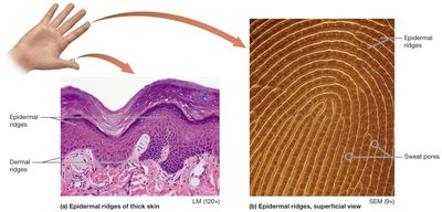
Skin markings are visible features resulting from the structure of the dermis and epidermis. Dermal ridges and epidermal ridges form fingerprints and enhance grip.
Dermal papillae: Form dermal ridges, which in turn create epidermal ridges.
Function: Increase friction and surface area for gripping.
Reticular Layer and Tension Lines
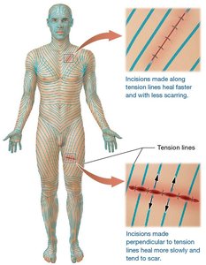
Tension lines (also called cleavage lines) are patterns formed by collagen fibers in the reticular layer. They are clinically important for surgical incisions.
Tension lines: Incisions made parallel to these lines heal faster and with less scarring.
Perpendicular incisions: Tend to heal more slowly and scar more.
Melanocytes and Skin Pigmentation
Melanocyte Function
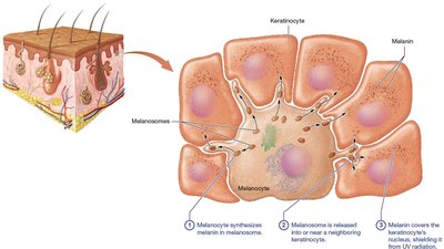
Melanocytes are specialized cells in the epidermis responsible for producing melanin, the pigment that gives skin its color and protects against UV radiation.
Melanin: Synthesized in melanosomes and transferred to keratinocytes.
Protection: Melanin covers the nucleus of keratinocytes, shielding DNA from UV damage.
Accessory Structures of the Skin
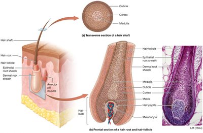
Hair
Hair is a keratinized structure produced by hair follicles in the dermis. It serves protective, sensory, and thermoregulatory functions.
Structure: Hair shaft, root, follicle, and bulb.
Layers: Cuticle, cortex, and medulla.
Associated structures: Arrector pili muscle, sebaceous glands, melanocytes.
Nails
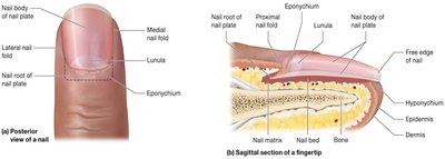
Nails are hard, keratinized plates covering the distal phalanges. They protect fingertips and enhance fine manipulation.
Parts: Nail body, nail root, lunula, nail matrix, nail bed, cuticle (eponychium), hyponychium.
Growth: Occurs from the nail matrix.
Sweat Glands
Sweat glands are exocrine glands involved in thermoregulation and waste excretion. There are several types:
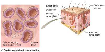
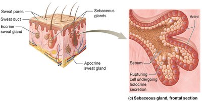
Eccrine glands: Most numerous, secrete watery sweat for cooling.
Apocrine glands: Found in specific areas (e.g., axilla), secrete thicker sweat, often associated with odor.
Ceruminous glands: Located in the ear canal, produce earwax.
Mammary glands: Specialized for milk production.
Eccrine Sweat Glands
Eccrine glands are distributed throughout the body and play a key role in temperature regulation.
Merocrine secretion: Sweat is released via exocytosis.
Structure: Coiled tubular glands opening directly onto skin surface.
Apocrine Sweat Glands
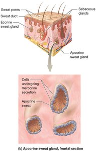
Apocrine glands are found in the axillary and genital regions, becoming active at puberty.
Secretion: Thicker, protein-rich sweat; may contribute to body odor.
Merocrine mechanism: Despite the name, apocrine glands use merocrine secretion.
Ceruminous and Mammary Glands
Ceruminous glands produce cerumen (earwax), which protects the ear canal. Mammary glands are modified sweat glands that produce milk.
Sebaceous Glands
Sebaceous glands secrete sebum, an oily substance that lubricates and waterproofs the skin and hair.
Holocrine secretion: Cells rupture to release sebum.
Location: Associated with hair follicles.
Clinical Aspects of the Integumentary System
Burns
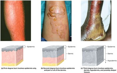
Burns are injuries to the skin caused by heat, chemicals, electricity, or radiation. They are classified by depth:
First-degree: Affect only the epidermis; redness and pain.
Second-degree: Involve epidermis and part of dermis; blistering.
Third-degree: Extend through dermis into deeper tissues; risk of infection and fluid loss.
Skin Cancer
Skin cancer is the uncontrolled growth of abnormal skin cells. The three main types are:
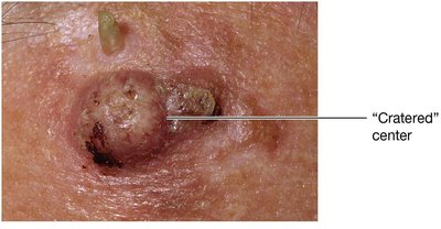
Basal Cell Carcinoma: Most common, arises from basal cells; rarely metastasizes.
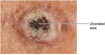
Squamous Cell Carcinoma: Originates from keratinocytes; may metastasize if untreated.
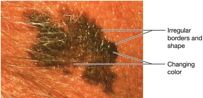
Malignant Melanoma: Arises from melanocytes; highly aggressive and prone to metastasis.
Basal Cell Carcinoma
Squamous Cell Carcinoma
Malignant Melanoma
Summary Table: Skin Glands
The following table summarizes the main types of skin glands and their characteristics:
Gland Type | Location | Secretion | Function |
|---|---|---|---|
Eccrine | Throughout body | Watery sweat | Thermoregulation |
Apocrine | Axilla, genital regions | Thicker, protein-rich sweat | Odor, stress response |
Ceruminous | Ear canal | Cerumen (earwax) | Protects ear canal |
Mammary | Breasts | Milk | Nourishment for infants |
Sebaceous | Associated with hair follicles | Sebum (oil) | Lubricates skin and hair |
Additional info: The notes above expand on brief points from the original material, providing definitions, examples, and clinical context for each structure and function of the integumentary system.
