 Back
The Integumentary System: Structure, Function, and Associated Features
Study Guide - Smart Notes
 Tailored notes based on your materials, expanded with key definitions, examples, and context.
The Integumentary System
Overview and Functions
The integumentary system, primarily composed of the skin, is the body's largest organ and serves as a protective covering. It performs multiple essential functions, including protection, temperature regulation, sensation, metabolic activities, blood storage, and waste elimination.
Protection: Keratinized cells form a physical barrier; skin secretions and melanin provide chemical protection.
Temperature Regulation: Sweat glands help dissipate heat.
Cutaneous Sensation: Skin contains tactile and pain receptors for sensory input.
Metabolic Functions: Skin synthesizes vitamin D precursor in response to UV radiation.
Blood Reservoir: Dermis can hold up to 5% of total blood volume.
Elimination of Wastes: Sweat excretes nitrogenous wastes, water, and NaCl.
Skin Structure
The skin consists of two main layers: the epidermis (outermost, protective, epithelial tissue) and the dermis (underlying, tough, dense connective tissue). Beneath the skin lies the superficial fascia, composed mainly of adipose tissue, which is not part of the skin itself.
Cells of the Epidermis
Keratinocytes: Most abundant; produce keratin, providing protection. Constantly replicate, replacing the epidermis every 25-45 days. Persistent friction leads to callous formation.
Melanocytes: Spider-shaped cells in the deepest epidermal layer; produce melanin pigment, which protects against UV damage.
Dendritic Cells (Langerhans cells): Star-shaped immune cells that ingest foreign substances and migrate to lymph nodes. Dermal macrophages also contribute to immune defense.
Tactile Epithelial Cells (Merkel cells): Located at the epidermis/dermis border; associated with sensory nerve receptors, enabling touch sensation.
Pigments of the Skin
Three main pigments contribute to skin color:
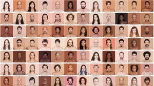
Melanin: Produced from tyrosine; ranges from reddish-yellow to brown-black. Production is influenced by genetics, hormones, age, inflammation, and UV exposure.
Carotene: Yellow-orange pigment from fruits and vegetables; accumulates in skin and converts to vitamin A, important for vision and skin health.
Hemoglobin: Iron-containing pigment in red blood cells; gives a pink hue to skin with low melanin due to visible capillaries.
Layers of the Epidermis
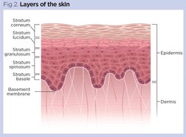
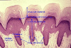
The epidermis is organized into five distinct layers, each with unique characteristics:
Stratum Basale: Deepest layer; actively produces keratinocytes and melanocytes.
Stratum Spinosum: Several cells thick; keratinocytes appear spiky due to desmosome connections and water loss.
Stratum Granulosum: 1-5 cells thick; keratinocytes begin filling with keratin.
Stratum Lucidum: Clear layer found only in thick skin (palms, soles); consists of a few rows of flat, dead keratinocytes.
Stratum Corneum: Outermost layer; 20-30 cells thick, composed of flattened, dead keratinocytes (dandruff, dry skin).
Thick vs. Thin Skin
Skin thickness varies by location:
Thick Skin: Found on palms, feet, and fingertips; includes the stratum lucidum.
Thin Skin: Covers most of the body; lacks the stratum lucidum.
Wrinkling of Fingers in Water
Wrinkling is a sympathetic response involving vasoconstriction in the dermis, leading to fluid shifts and skin puckering. This adaptation may improve grip in wet conditions.
Dermis
The dermis is a strong, flexible layer of dense connective tissue containing fibroblasts, macrophages, white blood cells, and mast cells. It is divided into two regions:
Papillary Dermis: Thin, loose areolar connective tissue; contains dermal papillae with capillary loops, pain receptors, and tactile corpuscles (Meissner's). Dermal ridges form fingerprints, aiding grip and sensation.
Reticular Dermis: Deepest and thickest layer; dense irregular connective tissue with thick collagen bundles, hair follicles, arrector pili muscles, and sweat glands.
Striae (Stretch Marks)
Extreme stretching of the skin tears the dermis, resulting in striae. Common causes include rapid growth, weight gain, pregnancy, and steroid use. Prevention strategies include hydration, moisturization, and massage.
Associated Skin Structures
Hair
Hair consists of flexible strands of dead, keratinized cells. It is thickest on the scalp, providing protection against trauma, heat loss, and UV radiation. Eyelashes and nasal hairs serve protective functions.
Regions: Root (bulb) and shaft, composed of medulla, cortex, and cuticle.
Color: Determined by melanin concentration; red hair contains pheomelanin, gray/white hair results from low melanin and air bubbles.
Types: Vellus (fine body hair) and terminal (scalp, eyebrows, lashes).
Growth Cycle: Varies by region; scalp hair grows actively for about 4 years, eyebrows for 2 months.
Baldness: Often genetically determined and linked to androgens.
Nails
Nails are clear, protective coverings of the dorsal distal fingers and toes, composed of hard keratin. They grow outward from the nail matrix, with the nail plate consisting of superficial keratinized cells and the nail bed rich in capillaries.
Clinical Indicators: Changes in nail appearance can signal health issues, such as onychomycosis (fungal infection), koilonychia (iron deficiency anemia), half-and-half nails (chronic kidney disease), Beau's lines (various illnesses), and yellow nail syndrome (chronic respiratory disease).
Sweat Glands
There are two main types of sweat glands:
Eccrine Glands: Numerous on hands, feet, and face; produce watery sweat containing NaCl, urea, and dermicidin. Sweat is acidic (pH 4-6).
Apocrine Glands: Located in axilla and anogenital areas; ducts open to hair follicles. Activated at puberty, producing odorous sweat with fatty and protein substances. Ceruminous and mammary glands are modified sweat glands.
Sebaceous Glands
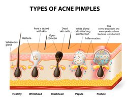
Holocrine glands that secrete sebum (oil) into hair follicles or onto the skin surface. Sebum lubricates hair and skin, prevents water loss, and has antibacterial properties.
Additional info:
Fingerprints are unique and genetically determined, aiding grip and enhancing tactile sensation.
Hair loss is multifactorial, involving thyroid function, age, immune dysfunction (alopecia), drugs, dietary deficiencies, genetics, stress, and pregnancy.
Eyebrows have evolved as a social tool, facilitating complex facial expressions and social interactions.
