 Back
BackThe Integumentary System: Structure, Function, and Healing
Study Guide - Smart Notes
 Tailored notes based on your materials, expanded with key definitions, examples, and context.
The Integumentary System
Introduction to the Integumentary System
The integumentary system consists of the skin and its accessory structures, including hair, nails, glands, and sensory receptors. It serves as a protective barrier between the internal and external environments and is the largest organ system by weight.
Organ Definition: A structure composed of two or more tissue types performing specific functions.
Accessory Structures: Hair, nails, glands, and sensory receptors.
Barrier Function: Protects against physical, chemical, and biological threats.
Skin and Its Tissues
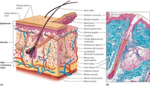
The skin is composed of two primary layers: the epidermis and dermis, separated by a basement membrane. Beneath these lies the subcutaneous (hypodermis) layer, which is not technically part of the skin but connects it to underlying tissues.
Epidermis: Thin, outer layer of stratified squamous epithelium; avascular.
Dermis: Thicker, inner layer with connective tissue, blood vessels, nerves, and muscle.
Basement Membrane: Anchors epidermis to dermis.
Subcutaneous Layer: Areolar and adipose tissue; insulates and contains major blood vessels.
The Epidermis
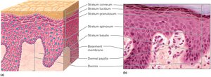
The epidermis is a stratified squamous epithelium lacking blood vessels. It consists of four layers in most areas and five in thick skin (palms, soles).
Stratum Basale: Deepest, dividing layer; nourished by dermal blood vessels.
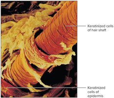
Keratinocytes: Cells that migrate outward, harden, dehydrate, and die (keratinization).
Stratum Spinosum, Granulosum, Corneum: Layers with increasing keratinization; stratum corneum is the outermost, composed of dead, flattened cells.
Stratum Lucidum: Found only in thick skin, between granulosum and corneum.
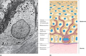
Melanocytes and Skin Color
Melanocytes are specialized cells in the deepest epidermal layer and dermis, producing melanin pigment. Melanin protects DNA from UV radiation and determines skin and hair color.
Melanin: Brownish-black (eumelanin) or reddish-yellow (pheomelanin).
Cytocrine Secretion: Transfer of melanin granules to nearby cells.
Skin Color: Determined by genetics, environment, and physiology; affected by melanin production, blood circulation, diet, and disease.
The Dermis
The dermis binds the epidermis to underlying tissues and contains connective tissue, blood vessels, nerve fibers, and accessory structures.
Dermal Papillae: Upward projections forming friction ridges (fingerprints).
Collagen and Elastic Fibers: Provide toughness and elasticity.
Blood Vessels: Supply nutrients and regulate temperature.
Accessory Structures: Hair follicles, glands, sensory receptors.
Accessory Structures of the Skin: Epidermal Derivatives
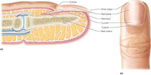
Nails
Nails are protective coverings over the ends of fingers and toes, consisting of a nail plate overlying the nail bed. The lunula is the visible, actively growing region.
Keratinization: Nails are harder than the stratum corneum.
Medical Diagnosis: Nail appearance can indicate health status.
Hair and Hair Follicles
Hair is found in most skin regions and develops from epithelial stem cells in hair follicles. The hair bulb at the follicle base contains the matrix, where cells divide and are nourished by dermal blood vessels.
Hair Shaft: Composed of dead, keratinized epithelial cells.
Hair Color: Determined by melanin type and amount; genetic lack of melanin causes albinism.
Arrector Pili Muscle: Smooth muscle causing goose bumps.
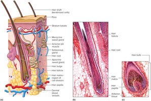
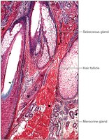
Skin Glands
Sebaceous Glands: Secrete sebum to waterproof and soften hair and skin; holocrine glands.
Sweat (Sudoriferous) Glands:
Eccrine: Respond to temperature; secrete onto skin surface.
Apocrine: Active at puberty; secrete into hair follicles; sweat contains proteins and fats.
Modified Sweat Glands: Ceruminous (ear wax) and mammary (milk).
Functions of the Skin
Skin Functions
The skin is essential for homeostasis, providing protection, waterproofing, UV defense, sensory reception, waste excretion, vitamin D synthesis, and temperature regulation.
Protection: Against pathogens, chemicals, and physical injury.
Waterproofing: Prevents water loss.
UV Protection: Melanin absorbs UV radiation.
Sensory Reception: Touch, pressure, pain, temperature.
Excretion: Removal of wastes via sweat.
Vitamin D Synthesis: Essential for bone and tooth development.
Temperature Regulation: Via sweat glands and blood vessel dilation/constriction.
Role of Skin in Body Temperature Regulation
The skin regulates body temperature through mechanisms controlled by the hypothalamus.
Heat Production: By active cells (heart, skeletal muscle, liver).
Heat Loss: Through radiation and sweat evaporation.
Skin’s Response to Temperature Changes
Hyperthermia (Excess Heat): Vasodilation and sweat gland activation.
Hypothermia (Excess Cooling): Vasoconstriction, sweat gland inactivation, shivering.
Healing of Wounds
Inflammation and Wound Healing
Inflammation is the body's response to injury, involving dilation and increased permeability of blood vessels. Healing depends on wound depth.
Superficial Cuts: Filled by reproducing epithelial cells.
Deep Wounds: Blood clot formation, scab development, fibroblast migration, collagen fiber production, debris removal, and tissue replacement.
Scar Formation: Excess collagen may form scars; large wounds may develop granulation tissue.
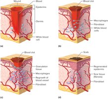
Table: Characteristics of Inflammation
Characteristic | Description |
|---|---|
Redness | Increased blood flow to the area |
Swelling | Increased permeability and fluid accumulation |
Heat | Increased blood flow and metabolic activity |
Pain | Release of chemicals and pressure on nerve endings |
Summary
The integumentary system is vital for protection, regulation, and sensation. Its complex structure and functions are essential for maintaining health and responding to injury. Understanding these components is crucial for students of anatomy and physiology.
