 Back
BackThe Integumentary System: Structure, Function, and Repair
Study Guide - Smart Notes
 Tailored notes based on your materials, expanded with key definitions, examples, and context.
Chapter 5: The Integumentary System
Overview of the Integumentary System
The integumentary system is the body's largest organ system, comprising the skin and its accessory structures. It serves as a protective barrier and plays vital roles in homeostasis, sensation, and metabolic processes.
Protection: Prevents fluid loss and protects against UV radiation (via melanin).
Excretion: Removes water, salts, and metabolic wastes through sweat.
Temperature Maintenance: Sweat glands regulate body temperature.
Nutrient Storage: Stores lipids in adipocytes within the dermis and hypodermis.
Vitamin D3 Synthesis: Epidermal cells produce vitamin D3, essential for calcium metabolism.
Sensory Detection: Contains receptors for touch, pressure, pain, and temperature.
The integumentary system accounts for approximately 16% of body weight and covers 1.5–2 m2 of surface area.
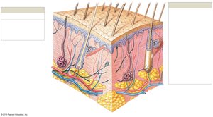
Components of the Integumentary System
Epidermis: Stratified squamous epithelium; avascular.
Dermis: Connective tissue; contains nerves, blood vessels, and sensory receptors.
Hypodermis: Subcutaneous layer; not technically part of the skin but supports it with adipose and areolar tissue.
Accessory Structures: Hair, nails, sebaceous (oil) glands, and sweat glands.
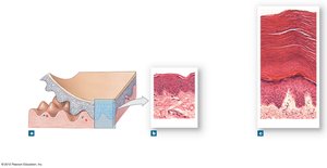
The Epidermis
Structure and Layers
The epidermis is composed primarily of keratinocytes and is organized into distinct layers (strata). It is avascular, relying on diffusion from the underlying dermis for nutrients.
Thin Skin: Four layers (strata); found on most of the body; ~0.08 mm thick.
Thick Skin: Five layers (includes stratum lucidum); found on palms and soles; ~0.5 mm thick.
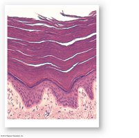
Strata of the Epidermis
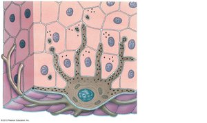
Stratum Basale: Deepest layer; single row of columnar keratinocytes attached to the basement membrane via hemidesmosomes. Contains stem cells, melanocytes (produce melanin), and tactile (Merkel) cells for sensation. Epidermal ridges interlock with dermal papillae, forming fingerprints.
Stratum Spinosum: 8–10 layers of keratinocytes connected by desmosomes. Contains dendritic (Langerhans) cells for immune defense. Last layer where cells divide.
Stratum Granulosum: 3–5 layers of keratinocytes; cells stop dividing and produce large amounts of keratin.
Stratum Lucidum: Present only in thick skin; flat, densely packed, keratinized cells.
Stratum Corneum: 15–30 layers of dead, fully keratinized cells; provides a tough, water-resistant barrier. Cells are shed after about two weeks.


Keratinization and Cornification
Keratinization: Process by which keratinocytes produce keratin, die, and become part of the protective outer layer.
Cornification: Synonymous with keratinization; results in the formation of the stratum corneum.
Exfoliation: Shedding of dead cells from the surface.
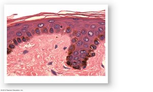
Epidermal Pigmentation
Skin color is primarily determined by the amount and distribution of melanin, produced by melanocytes in the stratum basale.
Melanin: Protects against UV radiation; production is stimulated by UV exposure.
Melanosomes: Vesicles that transfer melanin to keratinocytes.
Skin Color Variation: In fair skin, melanin is transferred only to the stratum basale and degrades quickly; in dark skin, transfer extends to the stratum granulosum, resulting in more persistent pigmentation.
Vitamin D3 Synthesis
When exposed to UV light, epidermal cells in the stratum spinosum and basale convert cholesterol into cholecalciferol (vitamin D3). The liver and kidneys further process this into calcitriol, which is essential for calcium and phosphorus absorption in the small intestine.
Deficiency: Leads to rickets in children (bone deformities).
Immune Function: Vitamin D is also important for immune system efficiency.
The Dermis and Hypodermis
Dermis Structure
The dermis lies beneath the epidermis and provides structural strength and elasticity to the skin. It is divided into two layers:
Papillary Layer: Areolar connective tissue; contains capillaries, lymphatics, and sensory neurons. Dermal papillae interlock with epidermal ridges.
Reticular Layer: Dense irregular connective tissue; contains collagen and elastic fibers, hair follicles, sweat and sebaceous glands, blood vessels, and nerves.
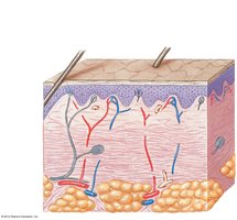
Hypodermis (Subcutaneous Layer)
Composed of areolar and adipose tissue.
Not technically part of the skin but anchors it to underlying tissues and organs.
Highly vascularized, serving as a site for fat storage and insulation.
Accessory Structures
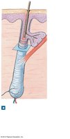
Hair and Hair Follicles
Hair is composed of keratinized cells and serves protective and sensory functions. Hair follicles are epidermal derivatives that extend into the dermis.
Functions: Protection from UV radiation, sensory input, and insulation (in other mammals).
Structure: Hair shaft, root, follicle, papilla (vascularized), arrector pili muscle (causes "goose bumps"), and associated sebaceous gland.
Nails
Composed of keratinized cells; protect the tips of fingers and toes.
Glands of the Integumentary System
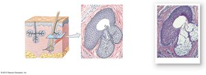
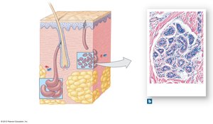
Sebaceous (Oil) Glands
These holocrine glands secrete sebum, an oily substance that lubricates and protects the skin and hair.
Location: Usually associated with hair follicles.
Function: Antibacterial properties and hair conditioning.
Control: Secretion is aided by the arrector pili muscle and regulated by the autonomic nervous system.
Sudoriferous (Sweat) Glands
Apocrine Sweat Glands: Actually use merocrine secretion; found in armpits, nipples, and pubic region. Secrete a cloudy, odorous fluid into hair follicles; function is likely vestigial. Begin functioning at puberty and are controlled by the endocrine and autonomic nervous systems.
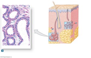
Merocrine (Eccrine) Sweat Glands: Discharge directly onto the skin surface; most numerous on palms and soles. Secrete a watery solution for thermoregulation (sensible perspiration), up to 4 L/hour.
Ceruminous Glands: Modified sweat glands in the external ear canal; produce cerumen (earwax).
Mammary Glands: Modified sweat glands that produce milk (not covered in detail here).
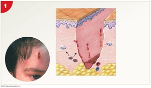
Integumentary Repair
Phases of Skin Repair
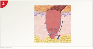
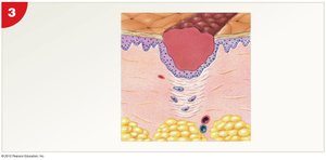
Skin repair involves a coordinated response between epithelial and connective tissue stem cells, immune cells, and fibroblasts.
Inflammatory Phase: Bleeding occurs, and mast cells trigger inflammation to increase blood flow and attract immune cells.
Migratory Phase: A scab forms (fibrin clot); cells from the stratum basale migrate to cover the wound. Phagocytic cells remove debris, and fibroblasts begin to produce collagen.
Proliferation Phase: Fibroblasts produce granulation tissue and new capillaries form. The scab is gradually dissolved.
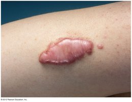
Scarring (Maturation) Phase: The epidermis is restored, but some scar tissue remains. Excessive collagen production can lead to keloids (raised scars).
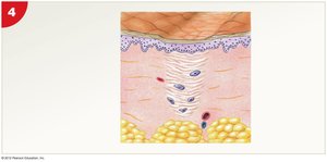
Keloid: An area of raised, thickened scar tissue resulting from overproduction of collagen during healing.
Summary Table: Layers of the Epidermis
Layer | Location | Key Features |
|---|---|---|
Stratum Corneum | Surface | 15–30 layers of dead, keratinized cells; water-resistant |
Stratum Lucidum | Thick skin only | Clear, flat, densely packed cells |
Stratum Granulosum | Below lucidum/corneum | 3–5 layers; keratin production; cells stop dividing |
Stratum Spinosum | Below granulosum | 8–10 layers; desmosomes; dendritic cells |
Stratum Basale | Deepest | Single layer; stem cells, melanocytes, Merkel cells |
