 Back
The Lumbar and Sacral Plexuses: Structure, Function, and Clinical Correlates
Study Guide - Smart Notes
 Tailored notes based on your materials, expanded with key definitions, examples, and context.
The Peripheral Nervous System: Nerve Plexuses
Overview of Peripheral Nerves and Plexuses
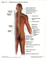
The peripheral nervous system (PNS) consists of nerves and ganglia outside the brain and spinal cord. Nerve plexuses are complex networks of intersecting nerves that primarily serve the limbs. The major plexuses include the cervical, brachial, lumbar, and sacral plexuses. These plexuses ensure that each limb muscle receives innervation from more than one spinal nerve, providing redundancy and protection against injury.
The Lumbar Plexus
Anatomy and Major Nerves of the Lumbar Plexus
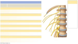
The lumbar plexus is formed by the ventral rami of spinal nerves T12–L4. It is located within the psoas major muscle and supplies the anterior and medial thigh, as well as parts of the abdominal wall and external genitalia.
Iliohypogastric nerve (T12–L1): Innervates the external and internal oblique and transverse abdominis muscles; provides sensory input from the skin over the inferior abdomen and buttocks.
Ilioinguinal nerve (L1): Supplies abdominal muscles and skin over the superior, medial thigh and portions of external genitalia.
Genitofemoral nerve (L1–L2): Sensory to skin over anteromedial thigh and portions of external genitalia.
Lateral femoral cutaneous nerve (L2–L3): Sensory to skin over anterior, lateral, and posterior thigh.
Femoral nerve (L2–L4): Motor to quadriceps, sartorius, pectineus, and iliopsoas muscles; sensory to anteromedial thigh and medial surface of the leg and foot.
Obturator nerve (L2–L4): Motor to gracilis and adductor muscles; sensory to medial thigh.
The Sacral Plexus
Anatomy and Major Nerves of the Sacral Plexus
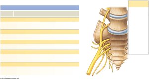
The sacral plexus is formed by the ventral rami of L4–S4, with a contribution from L4 and L5 (lumbosacral trunk). It is located on the posterior pelvic wall and supplies the posterior thigh, most of the lower leg, the entire foot, and part of the pelvis.
Superior gluteal nerve (L4–S1): Motor to gluteus minimus, gluteus medius, and tensor fasciae latae muscles.
Inferior gluteal nerve (L5–S2): Motor to gluteus maximus muscle.
Posterior femoral cutaneous nerve (S1–S3): Sensory to skin over perineum and posterior thigh and leg.
Sciatic nerve (L4–S3): Largest nerve in the body; branches into tibial and common fibular nerves; motor to hamstring muscles and adductor magnus; sensory and motor to most of the leg and foot.
Pudendal nerve (S2–S4): Motor to muscles of the perineum; sensory to skin over external genitalia.
Distribution of Major Nerves in the Lower Limb
Anterior and Posterior Views of Nerve Distribution
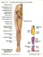
The lumbar and sacral plexuses give rise to nerves that innervate the lower limb. The femoral nerve supplies the anterior thigh, while the obturator nerve supplies the medial thigh. The sciatic nerve and its branches (tibial and common fibular nerves) supply the posterior thigh, leg, and foot.

Clinical Correlates: Nerve Injuries and Foot Deformities
Common Nerve Injuries Affecting the Lower Limb
Damage to specific nerves of the lumbar or sacral plexus can result in characteristic motor and sensory deficits, often leading to abnormal foot positions known as talipes (clubfoot) conditions.
Talipes Equinus (drop foot): Caused by damage to the common fibular nerve. The anterior muscles of the leg are paralyzed, preventing dorsiflexion of the foot.
Talipes Calcaneus (permanent dorsiflexion): Caused by damage to the tibial nerve. The calf muscles and posterior flexor muscles are paralyzed, preventing plantarflexion of the foot.
Other forms of talipes involve various combinations of abnormal foot positioning due to nerve or muscle dysfunction.

Summary Table: Major Nerves of the Lumbar and Sacral Plexuses
Nerve | Spinal Segments | Motor Distribution | Sensory Distribution |
|---|---|---|---|
Femoral | L2–L4 | Quadriceps, sartorius, pectineus, iliopsoas | Anteromedial thigh, medial leg and foot |
Obturator | L2–L4 | Gracilis, adductor magnus, brevis, longus | Medial thigh |
Sciatic | L4–S3 | Hamstrings, adductor magnus (part), all muscles below knee (via branches) | Most of leg and foot (via branches) |
Pudendal | S2–S4 | Muscles of perineum | External genitalia, perineal region |
Superior gluteal | L4–S1 | Gluteus medius, minimus, tensor fasciae latae | None (primarily motor) |
Inferior gluteal | L5–S2 | Gluteus maximus | None (primarily motor) |
Key Terms and Concepts
Plexus: A network of intersecting nerves.
Dorsiflexion: Upward movement of the foot at the ankle joint.
Plantarflexion: Downward movement of the foot at the ankle joint.
Talipes: A group of foot deformities, commonly known as clubfoot, resulting from abnormal muscle or nerve function.
