Back
BackThe Lymphatic and Immune Systems: Structure, Function, and Defense Mechanisms
Study Guide - Smart Notes
 Tailored notes based on your materials, expanded with key definitions, examples, and context.
The Lymphatic and Immune Systems
Overview and Functions
The lymphatic and immune systems are essential for maintaining fluid balance, absorbing dietary fats, and defending the body against pathogens. The lymphatic system collects interstitial fluid, filters it, and returns it to the bloodstream, while the immune system provides both innate and adaptive defenses against disease.
Immunity: Immune cells respond to foreign cells or chemicals, protecting the body from infection.
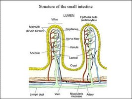
Lipid Absorption: Specialized lymphatic capillaries called lacteals absorb fats from the intestine and transport them to the bloodstream.
Fluid Recovery: The lymphatic system absorbs plasma proteins and excess fluid from tissues, returning them to the circulatory system.
Lymph and Lymphatic Capillaries
Structure and Function of Lymphatic Capillaries
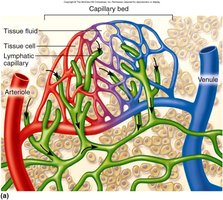
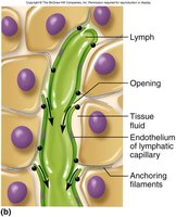
Lymphatic capillaries are microscopic, blind-ended vessels that collect lymph from interstitial spaces. Their unique structure allows for the uptake of large molecules, including proteins and pathogens.
Lymph: A clear, colorless fluid similar to plasma but with less protein.
Capillary Structure: Endothelial cells overlap loosely, forming valve-like flaps that open when interstitial fluid pressure is high and close when it is low.
Anchoring Filaments: Attach capillaries to surrounding tissues, preventing collapse.
Lacteals and Chyle
Lacteals are specialized lymphatic capillaries in the small intestine that absorb dietary fats, forming a milky fluid called chyle.
Lacteals: Transport absorbed fats into the circulatory system.
Chyle: Lymph mixed with emulsified fats, giving it a milky appearance.
Lymphatic Vessels and Lymph Flow
Structure of Lymphatic Vessels
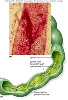
Lymphatic vessels transport lymph from capillaries to the circulatory system. They are structurally similar to veins but have thinner walls and more valves to prevent backflow.
Location: Often found superficial to blood vessels.
Valves: Ensure unidirectional flow of lymph.
Mechanisms of Lymph Flow
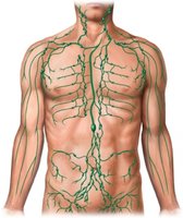
Lymph flows at low pressure and speed, aided by several mechanisms:
Skeletal muscle pump
Thoracic (respiratory) pump
Pulsation of nearby arteries
Smooth muscle contraction in vessel walls
Rapid blood flow in subclavian veins draws lymph in
Exercise increases lymphatic return
Route of Lymph Flow
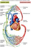
Lymph flows through a series of vessels and nodes before returning to the bloodstream:
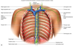
Lymphatic capillaries → Collecting vessels (pass through lymph nodes) → Lymphatic trunks → Collecting ducts (right lymphatic duct and thoracic duct) → Subclavian veins
Major Lymphatic Ducts
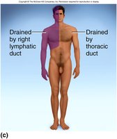
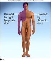
Right Lymphatic Duct: Drains lymph from the right arm, right side of head and thorax into the right subclavian vein.
Thoracic Duct: Larger and longer; begins as the cisterna chyli in the abdomen and drains lymph from the rest of the body into the left subclavian vein.
Lymphatic Disorders
Lymphedema
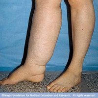
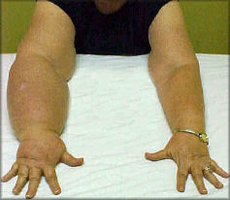
Lymphedema is localized fluid retention and tissue swelling caused by a compromised or blocked lymphatic system. It can lead to tissue damage and increased risk of infections.
Lymphatic Organs
Primary and Secondary Lymphatic Organs
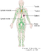
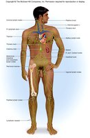
Lymphatic organs are classified as primary (where lymphocytes mature) or secondary (where immune responses are initiated).
Primary: Red bone marrow, thymus gland
Secondary: Lymph nodes, tonsils, spleen
Red Bone Marrow
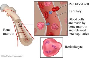
The site of hematopoiesis (blood cell formation). As blood cells mature, they enter the bloodstream by passing through the endothelium of blood vessels.
Thymus Gland
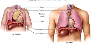
The thymus is a bilobed organ in the mediastinum with both lymphatic and endocrine functions. It is the site of T-cell development and atrophies after puberty.
T Lymphocytes: Mature in the thymus and are essential for adaptive immunity.
Lymph Nodes
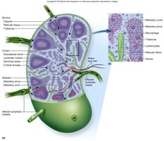
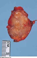
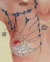
Lymph nodes are small, bean-shaped structures that filter lymph and are sites of B and T cell activation. They notify immune cells of pathogens and are common sites for metastatic cancer.
Structure: Cortex, medulla, afferent and efferent vessels, lymphatic nodules.
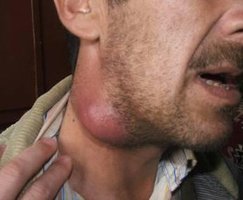
Lymphadenopathy: General term for lymph node diseases; lymphadenitis refers to swollen, painful nodes due to infection.
Tonsils
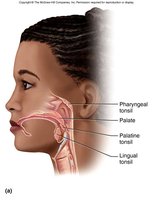
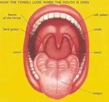
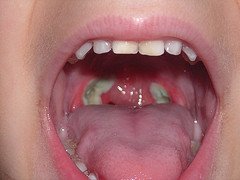
Tonsils are lymphatic tissues that protect against inhaled and ingested pathogens. There are three main types: palatine, lingual, and pharyngeal (adenoid).
Palatine Tonsils: Located at the back of the oral cavity; most often infected.
Lingual Tonsils: At the root of the tongue.
Pharyngeal Tonsil: On the wall of the pharynx.
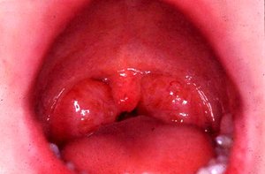
Spleen
The spleen filters blood, monitors for foreign antigens, and destroys aged red blood cells. It contains red pulp (erythrocyte-rich) and white pulp (lymphocyte-rich) regions. The spleen is highly vascular and vulnerable to injury.
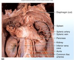
The Immune System
Defenses Against Pathogens
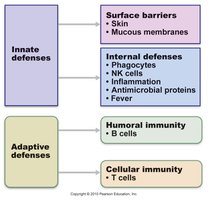
The immune system provides two main types of defense: innate (nonspecific) and adaptive (specific). Innate defenses are present from birth and respond broadly, while adaptive defenses develop after exposure to specific pathogens.
Innate Immunity: Includes surface barriers (skin, mucous membranes) and internal defenses (phagocytes, NK cells, inflammation, antimicrobial proteins, fever).
Adaptive Immunity: Involves B cells (humoral immunity) and T cells (cellular immunity).
Innate Immunity: First Line of Defense
Physical and chemical barriers prevent pathogen entry.
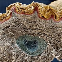
Skin: Keratinized cells form a tough barrier.
Mucous Membranes: Trap and expel pathogens.
Secretions: Lactic acid (sweat), lysozymes (saliva), sebum, gastric and vaginal secretions inhibit microbial growth.
Phagocytes
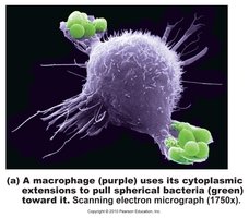
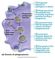
Phagocytes ingest and destroy pathogens. Neutrophils and macrophages are the main phagocytic cells.
Neutrophils: Patrol connective tissue, perform phagocytosis, and release toxic chemicals.
Macrophages: Derived from monocytes; specialized forms exist in various tissues (e.g., dendritic cells in skin, alveolar macrophages in lungs).
Natural Killer (NK) Cells
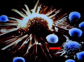
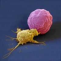
NK cells provide immune surveillance by targeting cancerous, virally infected, or otherwise abnormal cells.
Inflammation
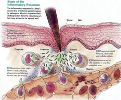
Inflammation is a localized response to infection or injury, characterized by pain, redness, immobility, swelling, and heat. It helps contain and eliminate pathogens and initiates tissue repair.
Pain: Caused by inflammatory chemicals and pressure on nerves.
Redness and Heat: Due to increased blood flow (hyperemia).
Swelling: Increased capillary permeability allows fluid to enter tissues.
Immobility: Loss of function may occur in severe inflammation.
Complement System
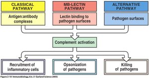
The complement system consists of over 30 plasma proteins that enhance both innate and adaptive immunity. They circulate in inactive form and are activated by pathogens via three pathways: classical, lectin, and alternative.
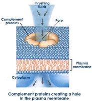
Functions: Opsonization (marking pathogens for phagocytosis), recruitment of inflammatory cells, and direct killing of pathogens via the membrane attack complex (MAC).
Fever (Pyrexia)
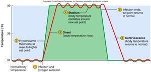
Fever is an abnormal elevation of body temperature, usually in response to infection. Pyrogens such as interleukin-1 are released by phagocytes and reset the hypothalamic thermostat. Fever increases metabolism, accelerates tissue repair, and inhibits pathogen reproduction.
Clinical Note: Low-grade fever may be beneficial, but temperatures above 105°F can cause delirium, and above 111°F may be fatal.