 Back
BackThe Lymphatic System and Immunity: Structure, Function, and Defense Mechanisms
Study Guide - Smart Notes
 Tailored notes based on your materials, expanded with key definitions, examples, and context.
The Lymphatic System and Immunity
Introduction to the Lymphatic System and Immunity
The lymphatic system is a critical component of the body's defense against disease. It consists of a network of vessels, tissues, and organs that help protect against pathogens—microscopic organisms such as viruses, bacteria, fungi, and parasites. The immune system, which includes the lymphatic system, provides immunity, or the ability to resist infection and disease, involving all body cells and tissues.
Pathogens: Disease-causing microorganisms that attack the body in specific ways.
Lymphocytes: White blood cells that identify, attack, and develop immunity to specific pathogens.
Immunity: The ability to resist infection and disease, involving both innate and adaptive mechanisms.

Components of the Lymphatic System
Main Components
The lymphatic system is composed of lymph, lymphatic vessels, lymphoid tissues and organs, and lymphoid cells. These components work together to maintain fluid balance, filter pathogens, and support immune responses.
Lymph: A fluid similar to plasma but lacking plasma proteins.
Lymphatic vessels: Transport lymph from peripheral tissues to the venous system.
Lymphoid tissues and organs: Include lymph nodes, tonsils, spleen, thymus, and MALT (mucosa-associated lymphoid tissue).
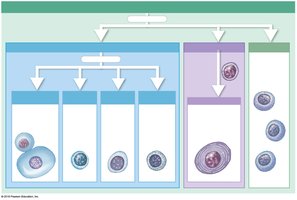
Lymphoid cells: Include lymphocytes (T cells, B cells, NK cells), phagocytes, and other immune cells.
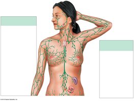
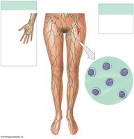
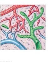
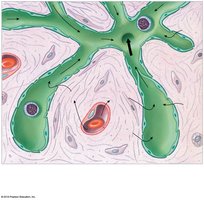
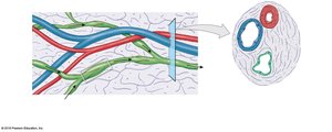
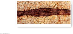
Lymphatic Capillaries and Vessels
Lymphatic capillaries are the entry points for interstitial fluid into the lymphatic system. They differ from blood capillaries in several ways, including being closed at one end, having larger diameters, thinner walls, and overlapping endothelial cells that act as one-way valves.
Lacteals: Specialized lymphatic capillaries in the small intestine that transport lipids.
One-way valves: Ensure unidirectional flow of lymph toward the venous system.
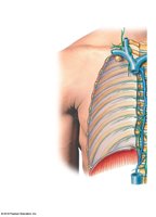
Lymphatic Ducts and Drainage
Lymphatic vessels converge to form larger trunks and ducts. The thoracic duct drains lymph from most of the body, while the right lymphatic duct drains the right upper quadrant. Both ducts empty into the subclavian veins.
Thoracic duct: Drains lymph from tissues inferior to the diaphragm and the left side of the upper body.
Right lymphatic duct: Drains lymph from the right upper body.
Lymphoid Tissues and Organs
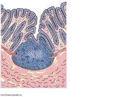
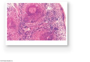
Lymphoid tissues are connective tissues dominated by lymphocytes. Lymphoid nodules are found in lymph nodes, spleen, tonsils, and along the digestive, urinary, and reproductive tracts. Major lymphoid organs include lymph nodes, thymus, and spleen.
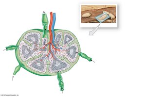
Lymph nodes: Filter lymph and remove antigens before returning fluid to circulation.
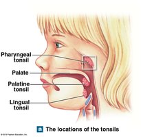
Tonsils: Five in the pharynx; protect against inhaled or ingested pathogens.
MALT: Lymphoid tissue associated with mucosal surfaces, including the appendix.
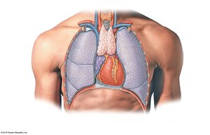
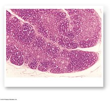
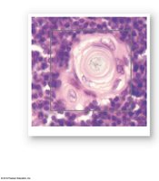
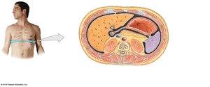
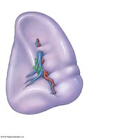
Thymus: Site of T cell maturation; atrophies after puberty.
Spleen: Filters blood, removes abnormal cells, stores iron, and initiates immune responses.
Innate and Adaptive Immunity
Types of Immunity
Immunity is the ability to resist and defend against infectious organisms and other harmful substances. There are two main types:
Innate (nonspecific) immunity: Present at birth, always works the same way, and defends against any type of invading agent.
Adaptive (specific) immunity: Develops after exposure to specific antigens and involves the activity of lymphocytes (T cells and B cells).
Innate Defenses
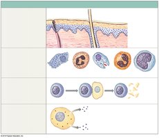
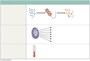
Innate defenses include physical barriers, phagocytes, immune surveillance, interferons, the complement system, inflammation, and fever.
Physical barriers: Skin and mucous membranes prevent pathogen entry.
Phagocytes: Engulf and destroy pathogens.
Immune surveillance: NK cells detect and destroy abnormal cells.
Interferons: Proteins that trigger antiviral defenses.
Complement system: Plasma proteins that enhance antibody action and cell lysis.
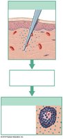
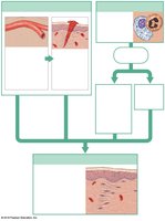
Inflammation: Localized response to injury or infection.
Fever: Elevated body temperature that accelerates defenses.
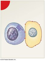
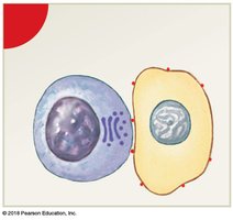
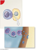
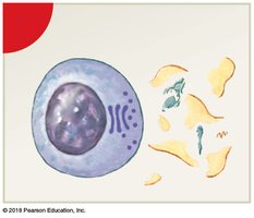
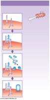
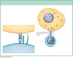
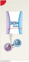
Immune Surveillance by NK Cells
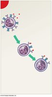
Natural killer (NK) cells identify and destroy abnormal cells by releasing perforins that create pores in the target cell membrane, leading to lysis.
Interferons and Complement System
Interferons are cytokines that trigger the production of antiviral proteins. The complement system consists of over 30 proteins that enhance the ability of antibodies and phagocytes to clear pathogens. Activation occurs via classical, lectin, or alternative pathways, leading to cell lysis, enhanced phagocytosis, and inflammation.
Inflammation and Fever
Inflammation is a localized response to tissue injury, characterized by redness, swelling, heat, and pain. It helps contain infection and initiates tissue repair. Fever is an elevation of body temperature induced by pyrogens, which enhances immune function and inhibits some pathogens.
Adaptive (Specific) Defenses
Overview of Adaptive Immunity
Adaptive immunity involves the coordinated activities of T cells and B cells. It is characterized by specificity, versatility, memory, and tolerance. Adaptive responses are triggered by antigens, which are chemical targets that stimulate immune responses.
Specificity: Each lymphocyte responds to a specific antigen.
Versatility: The body produces many types of lymphocytes.
Memory: Memory cells provide long-term immunity.
Tolerance: The immune system ignores self-antigens.
Forms of Immunity
Immunity can be classified as innate or adaptive, and as active or passive. Active immunity develops after exposure to antigens, while passive immunity is acquired by receiving antibodies from another source.
Active immunity: Can be naturally acquired (exposure to pathogens) or artificially acquired (vaccination).
Passive immunity: Can be naturally acquired (maternal antibodies) or artificially acquired (injection of antibodies).
T Cells and Cell-Mediated Immunity
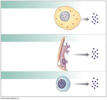
Antigen Presentation and MHC Proteins
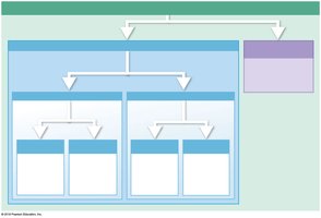
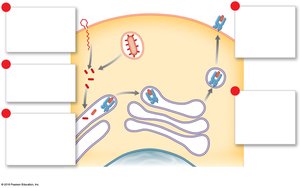
T cells are activated by antigens presented on the surface of cells by major histocompatibility complex (MHC) proteins. There are two classes:
Class I MHC: Found on all nucleated cells; present endogenous antigens to CD8 T cells (cytotoxic T cells).
Class II MHC: Found on antigen-presenting cells (APCs); present exogenous antigens to CD4 T cells (helper T cells).
Activation and Function of T Cells
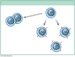
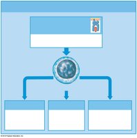
CD8 T cells differentiate into cytotoxic T cells, memory T cells, and regulatory T cells. CD4 T cells become helper T cells and memory helper T cells. Cytokines released by helper T cells stimulate both cell-mediated and antibody-mediated immunity.
B Cells and Antibody-Mediated Immunity
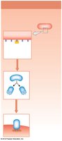
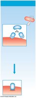
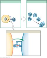
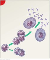
Activation of B Cells
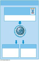
B cells are responsible for antibody-mediated immunity. They are activated when they encounter their specific antigen and receive costimulation from helper T cells. Activated B cells differentiate into plasma cells (which secrete antibodies) and memory B cells.
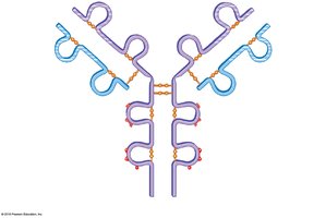
Antibody Structure and Classes
Antibodies (immunoglobulins) are Y-shaped proteins composed of two heavy and two light chains. The variable regions form antigen-binding sites, while the constant regions determine the antibody class (IgG, IgE, IgD, IgM, IgA).
IgG: Most abundant; crosses placenta; provides long-term immunity.
IgE: Involved in allergic responses and inflammation.
IgD: Functions in B cell sensitization.
IgM: First antibody produced in response to infection; causes agglutination.
IgA: Found in secretions; protects mucosal surfaces.
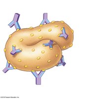
Antigen–Antibody Complex and Actions of Antibodies
Antibodies bind to antigens to form antigen–antibody complexes, leading to neutralization, precipitation, agglutination, activation of complement, attraction of phagocytes, opsonization, stimulation of inflammation, and prevention of pathogen adhesion.
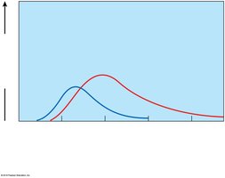
Primary and Secondary Immune Responses
The primary response to antigen exposure is slow and produces moderate antibody levels. The secondary response is faster and stronger due to memory cell activation, resulting in higher and more sustained antibody titers.
Immunocompetence and Integrated Immune Response
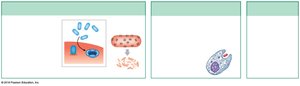
Immunocompetence
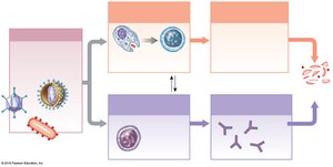
Immunocompetence is the ability to produce an immune response after exposure to an antigen. The immune response to bacterial infection involves both innate and adaptive mechanisms, including neutrophil and NK cell activity, cytokine release, antigen presentation, T and B cell activation, and antibody production.
Summary Table: Major Classes of Antibodies
Class | Main Function | Location/Notes |
|---|---|---|
IgG | Long-term immunity, crosses placenta | Most abundant in plasma |
IgE | Allergic response, inflammation | Bound to mast cells, basophils |
IgD | B cell sensitization | Surface of B cells |
IgM | First response, agglutination | Pentamer in plasma |
IgA | Mucosal immunity | Secretions (saliva, tears, mucus) |
