 Back
BackThe Lymphatic System and Immunity: Structure, Function, and Cellular Mechanisms
Study Guide - Smart Notes
 Tailored notes based on your materials, expanded with key definitions, examples, and context.
The Lymphatic System
Functions and Components
The lymphatic system is a network of organs, tissues, and vessels that works closely with the immune system to protect the body from pathogens, maintain fluid homeostasis, and absorb fats. It consists of blind-ended lymphatic vessels, lymphatic cells/tissue, and lymphoid organs such as tonsils, lymph nodes, spleen, and thymus.
Protection from Pathogens: Provides immune surveillance and response.
Fluid Homeostasis: Returns leaked fluids from blood vessels back to the bloodstream.
Absorption of Fats: Specialized capillaries called lacteals absorb dietary fats.
Lymphatic Vessels and Capillaries
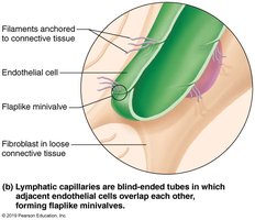
Lymphatic vessels transport lymph, a fluid composed of water, ions, and small molecules. Lymphatic capillaries are blind-ended tubes that weave between tissue cells and blood capillaries, allowing the uptake of larger molecules and particles that blood capillaries cannot.
Minivalves: Endothelial cells overlap to form one-way minivalves, anchored by collagen filaments, which respond to interstitial pressure.
Lacteals: Specialized capillaries in the intestinal mucosa absorb digested fats.
Lymph Transport
Lymph is transported through a low-pressure system, propelled by skeletal muscle contraction, thoracic pressure changes during breathing, valves, arterial pulsations, and smooth muscle contractions. Physical activity increases lymph flow, while immobilization helps retain inflammatory material for healing.
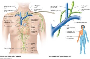
Main Lymphatic Trunks and Ducts
Lymphatic capillaries drain into larger collecting vessels, which pass through lymph nodes and eventually converge into lymphatic trunks and ducts. The thoracic duct and right lymphatic duct return lymph to the venous system.
Lymphatic Tissue and Organs
Lymphoid tissues house lymphocytes and macrophages, providing surveillance and immune response. They are composed largely of reticular connective tissue and are classified as nonencapsulated (e.g., MALT) or encapsulated (e.g., lymph nodes, spleen, thymus).
MALT: Mucosa-associated lymphoid tissue found beneath epithelium throughout the body, including Peyer's patches and BALT.
Lymphoid Follicles: Spherical bodies with germinal centers of proliferating B cells.
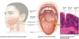
Tonsils
Tonsils are the simplest lymphoid organs, forming a ring of lymphatic tissue around the pharynx. They function to gather and remove pathogens in food or air and contain follicles with germinal centers.
Groups: Palatine ("tonsils"), pharyngeal ("adenoids"), lingual.
Lymph Nodes
Lymph nodes are bean-shaped structures found throughout the body, acting as lymph filters. They remove and destroy microorganisms and debris, preventing unwanted substances from entering the blood. Lymph nodes provide a site for lymphocyte activation and immune response.
Structure: External fibrous capsule, cortex (with follicles and germinal centers), medulla (with B cells, T cells, plasma cells).
Flow: Lymph enters via afferent vessels, passes through sinuses, and exits via efferent vessels at the hilum.
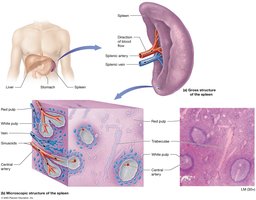
Spleen
The spleen is a blood-rich organ involved in immune function and blood filtration. It contains white pulp (immune function) and red pulp (breakdown of old blood cells).
White Pulp: Houses lymphocytes and macrophages targeting bloodborne pathogens.
Red Pulp: Cleanses blood of aged cells and platelets, stores breakdown products, platelets, and monocytes.
Thymus
The thymus is most active during childhood and is the site of T cell maturation. It contains a cortex with rapidly dividing T cells and a medulla with fewer lymphocytes. Regulatory T cells develop here to prevent autoimmunity.
The Immune System
Innate vs. Adaptive Immunity
The immune system is divided into innate (nonspecific) and adaptive (specific) immunity. Innate immunity provides immediate defense through physical barriers, chemical mediators, and certain leukocytes. Adaptive immunity is specific, systemic, and has memory, requiring priming by exposure to antigens.
Innate Immunity: Skin, mucous membranes, cilia, secretions, pH changes, microbiome.
Adaptive Immunity: Recognizes specific antigens, remembers previous encounters, and mounts stronger responses upon re-exposure.
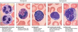
Leukocytes: Types and Functions
Leukocytes are grouped into granulocytes (neutrophils, eosinophils, basophils) and agranulocytes (lymphocytes, monocytes). Each type has distinct functions in immunity.
Neutrophils: Most numerous, phagocytic, "bacteria slayers."
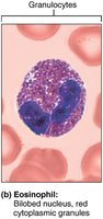
Eosinophils: Digestive enzymes, attack parasitic worms, involved in allergies and asthma.
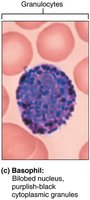
Basophils: Rarest, contain histamine, involved in inflammation and allergic reactions.
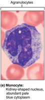
Monocytes: Differentiate into macrophages, phagocytic, present antigens to lymphocytes.
Lymphocytes: Include B cells, T cells, and natural killer cells.
Eosinophils
Basophils
Monocytes
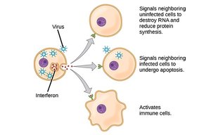
Innate Immunity: Complement, Cytokines, and Interferons
The complement system consists of 20 proteins that enhance antibody-mediated immunity, forming membrane attack complexes, stimulating phagocytosis, and promoting inflammation. Cytokines are proteins that enhance immune responses, including tumor necrosis factor and interleukins. Interferons are cytokines that prevent viral replication and activate immune cells.
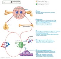
Origin and Development of Lymphocytes
B and T cells originate in red bone marrow and mature in primary lymphatic organs (B cells in bone marrow, T cells in thymus). In secondary lymphatic organs, lymphocytes interact with antigens and antigen-presenting cells to produce immune responses. Positive selection ensures survival of lymphocytes that react against antigens, while negative selection eliminates those that react against self-antigens.
Major Histocompatibility Complex (MHC)
MHC molecules are essential for antigen presentation and immune activation. Class I MHC is found on all nucleated cells and presents endogenous antigens, while Class II MHC is found on antigen-presenting cells and presents exogenous antigens.
T Cell Activation
T cell activation requires binding of the MHC class II/antigen complex to the T cell receptor and costimulation by cytokines or surface molecules. Proliferation occurs only if costimulation is present.
Helper and Cytotoxic T Cell Functions
Helper T Cells: Secrete cytokines to activate and enhance immune responses.
Cytotoxic T Cells: Kill cells with foreign MHC I antigens, including cancer cells and cells infected with intracellular pathogens.
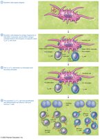
B Cell Activation and Antibody-Mediated Immunity
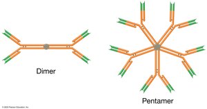
B cell activation requires antigen binding and costimulation by cytokines or surface molecules. Antibodies are Y-shaped proteins with constant and variable regions, effective against extracellular antigens.
Agglutination and Precipitation: Antibody binding creates clumps for easier phagocytosis.
Opsonization: Enhances targeting of pathogens.
Neutralization: Renders toxins inactive.
Stimulation of Inflammation: Draws immune cells to the area.
Primary vs. Secondary Immune Response
Primary immune response involves activation and proliferation of B cells, resulting in antibody production and memory B cells. Secondary response is faster and stronger due to memory B cells, often resulting in mild or no symptoms.
Acquired Immunity
Active Natural Immunity: Natural exposure to antigens.
Active Artificial Immunity: Vaccination.
Passive Natural Immunity: Transfer of antibodies from mother to fetus or baby.
Passive Artificial Immunity: Transfer of antibodies from immune animals.
Broader Connections
Understanding the lymphatic system and immunity is crucial for evaluating infection, immune disorders, and the effects of immunosuppressive therapies. The spleen's dual role in immune function and blood filtration highlights its importance in overall health, and damage to the spleen can impact both immunity and blood supply.
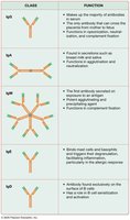
Summary Table: Antibody Classes and Functions
Class | Function |
|---|---|
IgG | Makes up the majority of antibodies in serum; crosses the placenta; neutralization and opsonization |
IgA | Found in secretions such as breast milk and saliva; protects mucosal surfaces |
IgM | First antibody secreted on exposure to an antigen; potent agglutinating and complement fixation |
IgE | Binds mast cells and basophils; triggers release of chemicals for allergic and inflammatory responses |
IgD | Antibody found exclusively on surface of B cells; helps activate B cells |
