 Back
BackThe Lymphatic System and Immunity: Structure, Function, and Clinical Relevance
Study Guide - Smart Notes
 Tailored notes based on your materials, expanded with key definitions, examples, and context.
The Lymphatic System
Overview and Main Functions
The lymphatic system is a network of vessels, nodes, and lymphoid tissues that plays a crucial role in maintaining fluid balance and defending the body against pathogens. It is essential for both the return of interstitial fluid to the bloodstream and the functioning of the immune system.
Maintaining Blood Volume and Composition: Collects excess interstitial fluid and returns it to the blood, preventing tissue swelling.
Immunity: Houses and transports lymphocytes, which are responsible for immune responses against pathogens, abnormal cells, and foreign proteins.
Components of the Lymphatic System
Lymph: Interstitial fluid that enters lymphatic vessels.
Lymphocytes: Primary cells of the lymphatic system, a type of leukocyte involved in immune responses.
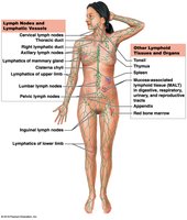
Lymphatic Vessels: Network of vessels that transport lymph from peripheral tissues back to the bloodstream.
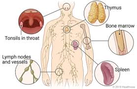
Lymphoid Tissues and Organs: Include lymph nodes, tonsils, MALT (mucosa-associated lymphoid tissue), appendix, spleen, red bone marrow, and thymus.
Lymphatic Vessels and Circulation
Structure and Function
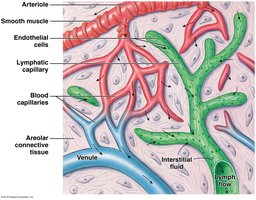
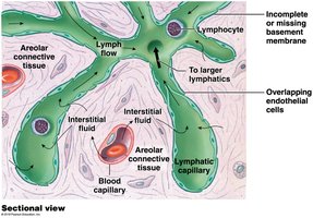
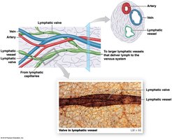
Lymphatic vessels begin as lymphatic capillaries in peripheral tissues and converge into larger vessels that return lymph to the venous system. They are closely associated with blood vessels and provide a one-way return trip for fluid.
Lymphatic Capillaries: Closed at one end, have larger diameters and thinner walls than blood capillaries, and feature overlapping endothelial cells that act as one-way valves.
Valves: Present in small to medium-sized lymphatic vessels to prevent backflow, especially in low-pressure environments.
Lacteals: Specialized lymphatic capillaries in the small intestine that transport absorbed lipids.
Major Lymphatic Ducts
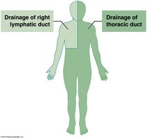
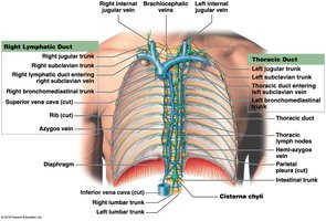
Thoracic Duct: Drains lymph from most of the body (below the diaphragm and left side above the diaphragm) into the left subclavian vein.
Right Lymphatic Duct: Drains lymph from the right side above the diaphragm into the right subclavian vein.
Cisterna Chyli: Expanded sac at the base of the thoracic duct that receives lymph from the lumbar and intestinal trunks.
Clinical Relevance: Lymphedema
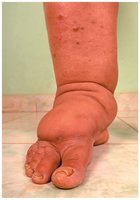
Lymphedema is swelling caused by blocked lymphatic drainage, leading to accumulation of interstitial fluid. It is most commonly seen in the limbs and can result in permanent swelling and increased risk of infection.
Lymphocytes and Immune Function
Classes of Lymphocytes
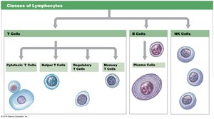
Lymphocytes are the main immune cells in the lymphatic system, making up 20–40% of circulating leukocytes. They are divided into three main classes:
T Cells (Thymus-derived): ~80% of lymphocytes; responsible for cell-mediated immunity.
B Cells (Bone marrow-derived): ~10–15%; responsible for antibody-mediated (humoral) immunity.
NK Cells (Natural Killer): ~5–10%; provide immune surveillance and destroy abnormal cells.
Lymphopoiesis
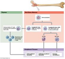
Lymphopoiesis is the production of lymphocytes, occurring primarily in the red bone marrow and thymus. Hematopoietic stem cells in the bone marrow give rise to all lymphocyte types. T cells mature in the thymus, while B cells and NK cells mature in the bone marrow and peripheral tissues.
Lymphoid Tissues and Organs
Lymphoid Nodules and MALT
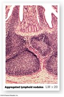
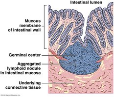
Lymphoid nodules are clusters of lymphocytes in areolar tissue, often found in mucosa-associated lymphoid tissue (MALT), which protects the epithelia of the digestive, respiratory, urinary, and reproductive tracts.
Tonsils
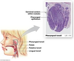
There are three pairs of tonsils (pharyngeal, palatine, and lingual) that form large lymphoid nodules in the pharynx and help protect against inhaled or ingested pathogens.
Lymph Nodes
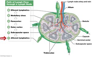
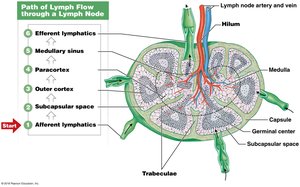
Lymph nodes are small, bean-shaped organs that filter lymph, removing pathogens before the fluid returns to the bloodstream. Lymph enters via afferent vessels, is filtered through the node, and exits via efferent vessels at the hilum.
Thymus
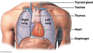
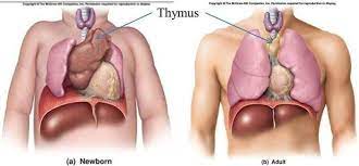
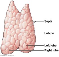
The thymus is a lymphoid organ in the mediastinum, posterior to the sternum. It is the site of T cell maturation and produces hormones called thymosins. The thymus is largest before puberty and shrinks with age.
Spleen
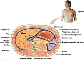
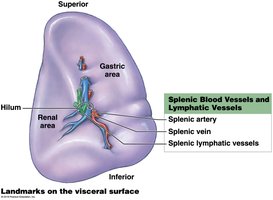
The spleen is the largest lymphoid organ, filtering blood, removing abnormal blood cells, and initiating immune responses to blood-borne antigens. It contains red pulp (rich in RBCs and macrophages) and white pulp (lymphoid tissue).
Immunity: Innate and Adaptive Defenses
Innate (Nonspecific) Immunity
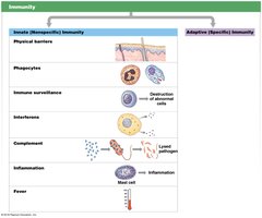
Innate immunity provides immediate, nonspecific defense against pathogens. It includes physical barriers, phagocytes, immune surveillance, interferons, complement system, inflammation, and fever.
Physical Barriers: Skin, mucous membranes, secretions, and hair prevent pathogen entry.
Phagocytes: Neutrophils, eosinophils, and macrophages engulf and destroy pathogens.
Immune Surveillance: NK cells monitor and destroy abnormal cells.
Interferons: Proteins that interfere with viral replication.
Complement System: Proteins that enhance antibody action and destroy pathogens.
Inflammation: Localized response to injury or infection.
Fever: Systemic response that inhibits pathogens and accelerates repair.
Adaptive (Specific) Immunity
Adaptive immunity is specific to particular antigens and involves T cells, B cells, and antibodies. It can be acquired naturally (infection) or artificially (vaccination), and is characterized by specificity, versatility, memory, and tolerance.
Active Immunity: Develops after exposure to antigen; long-term protection.
Passive Immunity: Antibodies received from another source; short-term protection.
Antigen Recognition and Immune Response
Antigen Presentation and MHC Proteins
Antigens are presented to T cells by MHC proteins on cell membranes. Class I MHC proteins are found on all nucleated cells and present endogenous antigens, while Class II MHC proteins are found on antigen-presenting cells and present exogenous antigens.
T Cell Activation and Differentiation
T cells are activated by antigen recognition and costimulation. They differentiate into cytotoxic T cells (destroy infected cells), memory T cells (provide long-term immunity), and regulatory T cells (modulate immune response).
B Cell Activation and Antibody Production
B cells require activation by helper T cells and differentiate into plasma cells (produce antibodies) and memory B cells (respond to future exposures).
Antibodies and Their Actions
Structure and Classes
Antibodies are Y-shaped proteins with variable regions for antigen binding. There are five main classes: IgG, IgE, IgD, IgM, and IgA, each with distinct roles in immune defense.
Primary and Secondary Responses
The primary response to antigen exposure is slow and limited, while the secondary response is rapid and robust due to memory cells.
Mechanisms of Antibody Action
Neutralization: Block pathogen binding sites.
Prevention of Pathogen Adhesion: IgA covers pathogens in secretions.
Activation of Complement: Enhances pathogen destruction.
Opsonization: Coating enhances phagocytosis.
Precipitation and Agglutination: Clumping of antigens for easier removal.
Clinical Considerations
Hypersensitivities and Allergies
Hypersensitivities are excessive immune responses to antigens (allergens), leading to inflammation and, in severe cases, anaphylaxis—a life-threatening systemic reaction.
Immune Disorders
Autoimmune Disorders: Immune system attacks self-antigens (e.g., multiple sclerosis, rheumatoid arthritis).
Immunodeficiency Diseases: Result from impaired immune function (e.g., AIDS caused by HIV infection).
Transplant Rejection: Immune response against transplanted tissues due to recognition of foreign MHC proteins.
Age-Related Changes
With age, the immune system becomes less effective due to thymic involution, reduced T and B cell responsiveness, and decreased immune surveillance, leading to increased susceptibility to infections and cancer.
Summary Table: Main Components and Functions of the Lymphatic System
Component | Structure | Main Function |
|---|---|---|
Lymph | Fluid in lymphatic vessels | Returns interstitial fluid to blood |
Lymphocytes | T cells, B cells, NK cells | Immune defense |
Lymphatic Vessels | Capillaries, vessels, ducts | Transport lymph |
Lymphoid Tissues | MALT, nodules | Immune surveillance |
Lymphoid Organs | Nodes, thymus, spleen | Filter lymph/blood, lymphocyte maturation |
