 Back
BackThe Lymphatic System and Immunity: Structure, Function, and Cellular Components
Study Guide - Smart Notes
 Tailored notes based on your materials, expanded with key definitions, examples, and context.
The Lymphatic System: Structure and Function
Overview of the Lymphatic System
The lymphatic system is a network of cells, tissues, and organs that defends the body against environmental pathogens, toxins, and abnormal body cells such as cancers. It is essential for maintaining fluid balance and supporting immune function.
Pathogens: Microscopic organisms that cause disease, including viruses, bacteria, fungi, and parasites. Each pathogen attacks the body in a specific way.
Immunity: The ability to resist infections or disease, involving all body cells and tissues, not just those of the lymphatic system.
Lymphocytes: Specialized white blood cells that identify, attack, and develop immunity to specific pathogens.
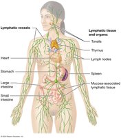
Components of the Lymphatic System
Lymph: A fluid similar to plasma but with fewer proteins, originating as interstitial fluid (IF).
Lymphatic vessels: Transport lymph from peripheral tissues to the venous system.
Lymphoid tissues: High concentrations of lymphocytes and macrophages, not enclosed by a capsule.
Lymphoid organs: High concentrations of lymphocytes and macrophages, enclosed in a capsule (e.g., lymph nodes, spleen, thymus).
Functions of the Lymphatic System
Lymphocyte production: Occurs in lymphoid tissues (e.g., tonsils), lymphoid organs (e.g., spleen, thymus), and red bone marrow. T-cells mature in lymphatic tissues and organs and migrate to sites of injury or infection.
Drainage of excess interstitial fluid: Returns fluid to the blood, maintaining blood volume and ensuring similar composition of interstitial fluid throughout the body.
Immunity: Lymph passes through lymph nodes, where immune cells can activate an immune response if needed.
Lymphatic Vessels and Circulation
Lymphatic Capillaries and Vessels
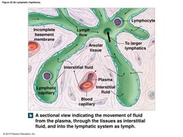
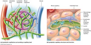
Lymphatic vessels begin as small, blind-ended lymphatic capillaries that differ from blood capillaries in several ways:
Start as pockets rather than tubes
Have larger diameters and thinner walls
Have an irregular outline
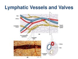
Endothelial cells overlap, forming one-way valves that allow entry of fluids, solutes, viruses, and bacteria, but prevent their return to interstitial fluid
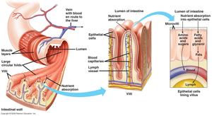
Lacteals
Lacteals are specialized lymphatic capillaries in the small intestine that transport lipids from the digestive tract.
Lymphatic Vessel Structure
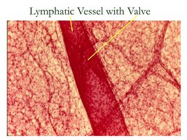
Lymph flows from lymphatic capillaries to larger lymphatic vessels, which contain one-way valves and travel alongside veins toward the trunk.
Lymph Circulation Pathways
Superficial lymphatics: Found in the skin, mucous membranes, and serous membranes lining body cavities.
Deep lymphatics: Larger vessels accompanying deep arteries and veins, found in the neck, limbs, and trunk.
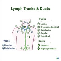
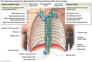
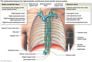
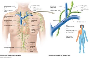
Superficial and deep lymphatics join to form lymphatic trunks, which empty into two major collecting vessels: the thoracic duct and the right lymphatic duct.
Major Lymphatic Ducts
Right lymphatic duct: Collects lymph from the right jugular, right subclavian, and right bronchomediastinal trunks; empties into the right subclavian vein.
Thoracic duct: Begins at the cisterna chyli, receives lymph from the right and left lumbar trunks and the intestinal trunk, and collects lymph from the left bronchomediastinal, left subclavian, and left jugular trunks; empties into the left subclavian vein.
Lymphoid Tissues and Organs
Lymphoid Tissue and Nodules
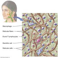
Lymphoid tissue consists of loose reticular connective tissue dominated by lymphocytes and thin reticular fibers that trap pathogens. Lymphoid nodules are areolar tissue with densely packed dividing lymphocytes and do not have a fibrous capsule.
Distribution of Lymphoid Nodules
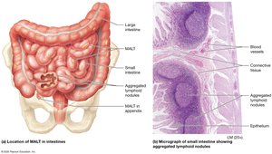
Found in lymph nodes, spleen, respiratory tract (tonsils), and along digestive, urinary, and reproductive tracts.
Mucosa-Associated Lymphatic Tissue (MALT): Collection of unencapsulated lymphoid tissue associated with the digestive system, including Peyer's patches and the appendix.
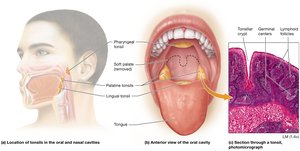
Tonsils: Large nodules in the walls of the pharynx.
Lymphoid Organs
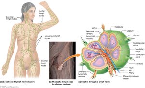
Lymph nodes: Small clusters of lymphatic tissue located along lymphatic vessels, concentrated in the neck, armpit, and groin. They filter lymph before returning it to venous circulation and stop pathogens before they reach vital organs.
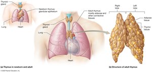
Thymus: Located in the mediastinum, large and active in infants and children, atrophies after puberty. Secretes thymosin to stimulate stem cell divisions and T cell differentiation.
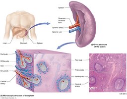
Spleen: Largest lymphoid organ, located in the left upper quadrant. Functions include removal of abnormal red blood cells, initiation of immune responses, and storage of red blood cells and platelets.
Cells and Proteins of the Innate and Adaptive Immune Systems
Body Defenses: Innate and Adaptive Immunity
The body uses two categories of defenses to resist infection, illness, and disease:
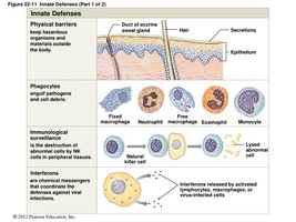
Innate (nonspecific) defenses: Provide general protection against any type of invading agent.
Adaptive (specific) defenses: Protect against specific pathogens and depend on the activity of lymphocytes. Develops after exposure to environmental hazards.
Cells of Innate Immunity
Phagocytes: Engulf and degrade cellular debris, foreign compounds, and pathogens. Two classes: Microphages (neutrophils and eosinophils) and Macrophages (derived from monocytes).
Basophils: Aid in the mobility and action of other white blood cells by releasing leukotrienes, histamine, and heparin.
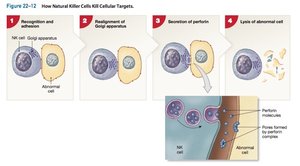
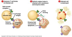
Natural Killer (NK) cells: Attack foreign cells, virus-infected cells, and cancer cells by releasing perforin, which perforates the plasma membrane of the target cell, leading to cytolysis.

Cells of Adaptive Immunity
Lymphocytes: Make up 20-30% of circulating leukocytes. Types include:
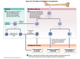
T cells: Thymus-dependent, provide cell-mediated immunity, attack foreign cells or cells infected by viruses.
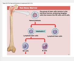
B cells: Bone marrow-derived, provide antibody-mediated immunity, attack antigens by producing specific antibodies.
NK cells: Natural killer cells, part of both innate and adaptive immunity.
Lymphopoiesis: Production of lymphocytes in red bone marrow, thymus gland, and peripheral lymphoid tissues.
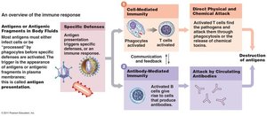
Functional Types of T and B Cells
Cytotoxic T cells: Attack cells infected by viruses, responsible for cell-mediated immunity, release perforin to destroy plasma membranes.
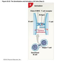
Helper T cells: Stimulate proliferation and differentiation of T cells and B cells.
B cells: Do not directly attack invaders; instead, they produce antibodies. Activated B cells divide into plasma cells, which secrete antibodies.
Additional info: The lymphatic system is essential for both fluid balance and immune defense. Its vessels, tissues, and organs work together to filter lymph, produce immune cells, and mount responses to pathogens. Understanding the structure and function of the lymphatic system is foundational for comprehending the body's defense mechanisms.
