 Back
BackThe Lymphatic System and Immunity: Structure, Function, and Clinical Relevance
Study Guide - Smart Notes
 Tailored notes based on your materials, expanded with key definitions, examples, and context.
The Lymphatic System and Immunity
Introduction to the Lymphatic and Immune Systems
The lymphatic and immune systems work together to protect the body from cellular injury, pathogens, and disease. The lymphatic system is a one-way network that returns excess interstitial fluid to the cardiovascular system and houses immune cells. The immune system, composed of cells and proteins, provides defense against foreign invaders through innate and adaptive mechanisms.
Lymphatic system: Includes lymphatic vessels (blind-ended tubes) and lymphatic tissues/organs (tonsils, lymph nodes, spleen, thymus).
Immune system: Consists of leukocytes (white blood cells) and plasma proteins, including T cells (from thymus) and B cells (from bone marrow).

Functions of the Lymphatic System
The lymphatic system performs three main functions essential for homeostasis and immunity:
Regulation of interstitial fluid volume: Returns 2–4 liters/day of fluid lost from plasma to circulation, preventing edema and maintaining blood pressure.
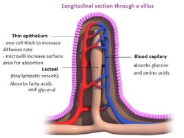
Absorption of dietary fats: Fats too large for blood capillaries enter lymphatic vessels (lacteals) in the small intestine and are transported with lymph.
Immune functions: Lymphoid organs filter pathogens and house leukocytes for immune surveillance and maturation.
Lymphatic Vessels and Lymph Circulation
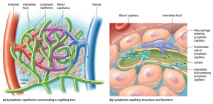
Lymphatic vessels collect and transport lymph through a low-pressure system aided by valves and smooth muscle contractions. Lymphatic capillaries are highly permeable, allowing entry of fluid, proteins, and immune cells.
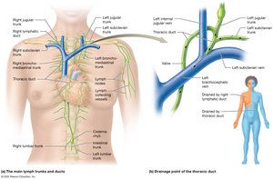
Lymph-collecting vessels merge into nine main lymph trunks, draining specific body regions.
Cisterna chyli: Receives lymph from intestinal and lumbar trunks, draining into the thoracic duct (left side of body).
Right lymphatic duct: Drains upper right side of body into the right subclavian vein.
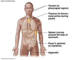
Lymphoid Tissues and Organs
Lymphoid organs provide sites for immune cell residence, activation, and pathogen filtration. Major lymphoid organs include:
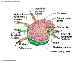
Lymph nodes: Filter lymph, trap pathogens, and contain lymphocytes and macrophages.
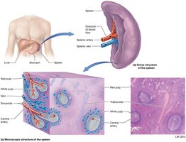
Spleen: Filters blood, removes old erythrocytes, and supports immune surveillance.
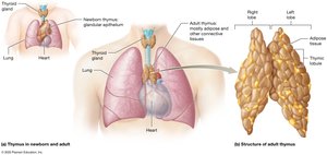
Thymus: Site of T cell maturation; most active in youth, atrophies with age.
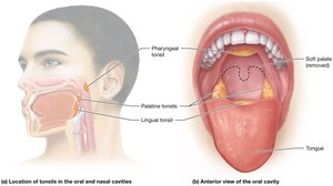
Tonsils: Trap pathogens entering through the oral and nasal cavities.
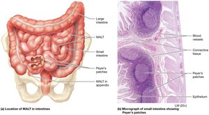
Peyer’s patches and appendix: Protect the gastrointestinal tract from pathogens.
MALT (Mucosa-associated lymphoid tissue): Includes tonsils, Peyer’s patches, and appendix, guarding mucosal surfaces.
Immunity: Body Defenses
Lines of Defense
The immune system provides three lines of defense against pathogens:
First line: Surface barriers (skin and mucous membranes) block pathogen entry.
Second line: Innate immunity—cells and proteins provide rapid, nonspecific defense.
Third line: Adaptive immunity—lymphocytes and antibodies provide specific, long-term protection.

Innate (Nonspecific) Immunity
Innate immunity responds to all pathogens in the same way and is the dominant response in the first 12 hours after exposure. It includes:
Surface barriers: Skin (keratinized, acidic sebum) and mucous membranes (mucus, acid) deter pathogen entry.
Phagocytic cells: Macrophages, neutrophils, dendritic cells, and eosinophils ingest and destroy pathogens.
Nonphagocytic cells: Natural killer (NK) cells, mast cells, and basophils mediate inflammation and cytotoxicity.
Antimicrobial proteins: Complement proteins and cytokines enhance immune responses.
Inflammation and fever: Local and systemic responses to infection or injury.

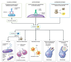
The Complement System
The complement system consists of over 20 plasma proteins that enhance both innate and adaptive immunity. They are activated via three pathways (classical, lectin, alternative) and mediate pathogen destruction, opsonization, inflammation, and cell lysis.
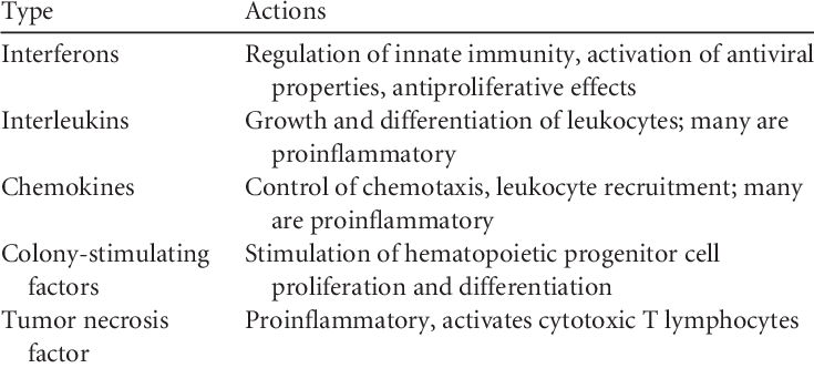
Cytokines
Cytokines are proteins produced by immune cells that regulate and enhance immune responses, including cell recruitment, inflammation, and activation of lymphocytes.
Type | Actions |
|---|---|
Interferons | Regulation of innate immunity, activation of antiviral properties, antiproliferative effects |
Interleukins | Growth and differentiation of leukocytes; many are proinflammatory |
Chemokines | Control of chemotaxis; leukocyte recruitment; many are proinflammatory |
Colony-stimulating factors | Stimulation of hematopoietic progenitor cell proliferation and differentiation |
Tumor necrosis factor | Proinflammatory; activates cytotoxic T lymphocytes |
Inflammatory Response and Fever
Inflammation is a local innate response to injury or infection, characterized by redness, swelling, heat, and pain. Fever is a systemic response, raising body temperature to enhance immune function and inhibit pathogens.
NSAIDs and corticosteroids: Medications that reduce inflammation by inhibiting prostaglandin and leukotriene synthesis.
Antipyretics: Drugs that lower fever by resetting the hypothalamic thermostat.
Adaptive (Specific) Immunity
Overview of Adaptive Immunity
Adaptive immunity is specific to particular antigens and has memory, allowing for a faster and stronger response upon re-exposure. It is divided into two arms:
Cell-mediated immunity: Involves T cells (helper and cytotoxic) that target infected or abnormal cells.
Humoral (antibody-mediated) immunity: Involves B cells that produce antibodies targeting extracellular pathogens.

Cell-Mediated Immunity
T cells recognize antigens presented by MHC molecules. Cytotoxic T cells (CD8+) kill infected or abnormal cells, while helper T cells (CD4+) secrete cytokines to enhance immune responses.
Antigen: Any substance recognized by B or T cells; immunogens elicit an immune response.
MHC molecules: Class I (all nucleated cells, present endogenous antigens to CD8+ T cells); Class II (APCs, present exogenous antigens to CD4+ T cells).
T cell activation: Requires antigen presentation, costimulation, and cytokine signaling.
Humoral (Antibody-Mediated) Immunity
B cells recognize antigens, become activated (with T cell help), and differentiate into plasma cells (secreting antibodies) and memory B cells. Antibodies (immunoglobulins) neutralize, opsonize, and agglutinate pathogens.
Antibody structure: Y-shaped molecule with variable (antigen-binding) and constant regions; five classes (IgG, IgA, IgM, IgE, IgD).
Immunological memory: Memory B cells enable rapid secondary responses.
Vaccination: Induces primary immune response and memory cell formation for future protection.

Types of Immunity
Active immunity: Results from direct exposure to antigen (infection or vaccination); long-lasting due to memory cell formation.
Passive immunity: Transfer of antibodies from another source (e.g., maternal antibodies, antibody therapy); temporary protection.
Disorders of the Immune System
Hypersensitivity Disorders
Hypersensitivity disorders are overreactions of the immune system that damage tissues. Four types are classified by mechanism:
Type I (Immediate): Allergies, anaphylaxis; mediated by IgE and mast cell degranulation.
Type II (Antibody-mediated): Antibodies target self-antigens (e.g., transfusion reactions, some drug allergies).
Type III (Immune complex-mediated): Immune complexes deposit in tissues, causing inflammation and damage.
Type IV (Delayed-type): T cell-mediated; includes contact dermatitis and tuberculin skin test reactions.
Immunodeficiency Disorders
Immunodeficiency disorders result from failure of one or more immune components:
Primary: Genetic or developmental defects (e.g., SCID, hypogammaglobulinemia).
Secondary: Acquired (e.g., HIV/AIDS, cancer, immunosuppressive therapy).
Autoimmune Disorders
Autoimmune disorders occur when the immune system attacks self-antigens, leading to tissue damage. Examples include multiple sclerosis, type 1 diabetes mellitus, systemic lupus erythematosus, and rheumatic fever.
Summary Table: Key Components of the Immune System
Cell Type | Description | Function |
|---|---|---|
Neutrophil | Most abundant granulocyte | Phagocytosis, first responder to infection |
Macrophage | Derived from monocytes | Phagocytosis, antigen presentation |
Dendritic cell | APC in tissues | Antigen presentation to T cells |
Eosinophil | Granulocyte | Defense against parasites |
Basophil/Mast cell | Granulocyte | Release histamine, mediate inflammation |
NK cell | Lymphocyte | Cytotoxicity against infected/cancerous cells |
B cell | Lymphocyte | Antibody production |
T cell | Lymphocyte | Cell-mediated immunity |
Additional info: This guide integrates textbook, lecture, and handwritten notes to provide a comprehensive overview of the lymphatic system and immunity for ANP college students.
