 Back
BackThe Lymphatic System and Immunity: Structure, Function, and Clinical Relevance
Study Guide - Smart Notes
 Tailored notes based on your materials, expanded with key definitions, examples, and context.
The Lymphatic System and Immunity
Introduction to the Lymphatic and Immune Systems
The lymphatic and immune systems work together to protect the body from cellular injury and pathogens. The lymphatic system consists of a network of vessels, tissues, and organs that maintain fluid balance, absorb dietary fats, and support immune functions. The immune system defends against internal and external threats through a coordinated response involving various cell types and molecules.
Functions of the Lymphatic System
Regulation of Interstitial Fluid Volume
Interstitial fluid is the fluid that surrounds tissue cells. Blood capillaries lose about 2–4 liters of fluid per day to the interstitial space due to filtration pressure.
Lymphatic vessels collect this excess fluid, now called lymph, and return it to the cardiovascular system, maintaining blood volume and pressure.
Absorption of Dietary Fats
Dietary fats are too large to enter blood capillaries directly. Instead, they are absorbed by specialized lymphatic capillaries called lacteals in the small intestine.
These fats travel through the lymphatic system and are eventually delivered to the bloodstream.
Immune Functions
Lymphoid organs filter pathogens from lymph and blood, house leukocytes, and support their maturation.
Lymphatic Vessels and Lymph Circulation
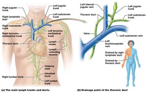
Main Lymphatic Trunks and Ducts
Lymphatic vessels begin as blind-ended capillaries in tissues, forming a one-way system that moves lymph away from tissues. These vessels merge into larger lymph trunks and ducts, which drain lymph from specific body regions.
Right and Left Lumbar trunks: Drain lower limbs and pelvic area.
Jugular trunks: Drain head and neck.
Intestinal trunk: Receives fat-containing lymph from the small intestine.
Bronchomediastinal trunks: Drain thoracic cavity.
Subclavian trunks: Drain upper limbs.
Cisterna chyli: Large vessel receiving lymph from lumbar and intestinal trunks.
Thoracic duct: Drains lower body and left upper body into the left subclavian vein.
Right lymphatic duct: Drains upper right side of the body into the right subclavian vein.
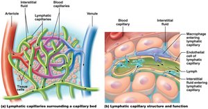
Structure and Function of Lymphatic Capillaries
Lymphatic capillaries form a weblike network around blood capillary beds and are structurally distinct from blood capillaries.
They are blind-ended, allowing only one-way movement of lymph.
Endothelial cells of lymphatic capillaries are not tightly joined, enabling them to open and close in response to interstitial fluid pressure, allowing large volumes of fluid and even cells to enter.
Lacteals are specialized lymphatic capillaries in the small intestine for fat absorption.
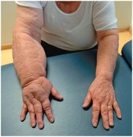
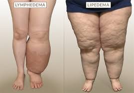
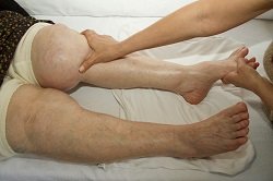
Clinical Relevance: Lymphedema
Definition and Causes
Lymphedema is severe swelling due to accumulation of interstitial fluid, often resulting from surgical removal or blockage of lymphatic vessels (e.g., by parasites).
Prevents proper return of lymph to the cardiovascular system, causing tissue enlargement and potential disfigurement.
Lymphoid Tissues and Organs
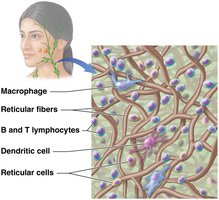
Reticular Tissue and Cell Types
Lymphatic system is composed of reticular tissue, a loose connective tissue with reticular fibers that trap pathogens.
Key cell types: Macrophages (phagocytes), B and T lymphocytes (immune cells), Dendritic cells (antigen-presenting), and Reticular cells (produce reticular fibers).
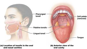
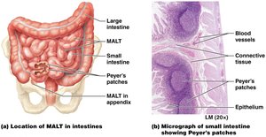
Mucosa-Associated Lymphatic Tissue (MALT)
MALT protects mucous membranes exposed to pathogens, found in the oral/nasal cavities, gastrointestinal tract, respiratory passages, and genitourinary tract.
Specialized MALT includes tonsils, Peyer's patches (in the ileum), and the appendix.
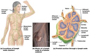
Lymph Nodes
Small, bean-shaped clusters located along lymphatic vessels; filter lymph and trap pathogens.
Major clusters: axillary, cervical, inguinal, and mesenteric lymph nodes.
Structure: Cortex (lymphoid follicles, T cells), Medulla (mature B cells), and reticular network for filtration.
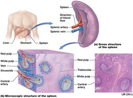
Spleen
Largest lymphoid organ, located in the left upper quadrant of the abdomen.
Red pulp: destroys old erythrocytes; White pulp: filters pathogens and contains immune cells.
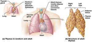
Thymus
Located in the superior mediastinum; site of T cell maturation.
Large and active in children, atrophies with age, replaced by fat in adults.
Contains cortex (densely packed T cells) and medulla (site of destruction of self-reactive T cells).
Overview of the Immune System
Lines of Defense
First line of defense: Surface barriers (skin, mucous membranes) block pathogen entry.
Second line of defense: Innate immunity (cells and proteins) responds quickly and non-specifically to pathogens.
Third line of defense: Adaptive immunity (B and T cells) responds specifically to antigens and has memory.
Types of Immunity
Innate (nonspecific) immunity: Rapid, general response to all pathogens; includes phagocytes, antimicrobial proteins, and inflammation.
Adaptive (specific) immunity: Slower, antigen-specific response; includes cell-mediated (T cells) and antibody-mediated (B cells) immunity; has immunological memory.
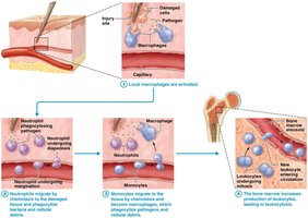
Cells of Innate Immunity
Phagocytic Cells
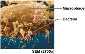
Macrophages: Mature from monocytes, highly active phagocytes.
Neutrophils: Most active phagocytes, especially against bacteria.
Eosinophils: Target parasites and participate in allergic responses.
Dendritic cells: Present antigens to T cells, activating adaptive immunity.
Nonphagocytic Cells
Natural Killer (NK) cells: Destroy infected or abnormal cells.
Basophils and Mast cells: Release inflammatory mediators, especially in allergic responses.
The Inflammatory Response
Stages of Inflammation
Damaged cells release inflammatory mediators, causing local changes (vasodilation, increased permeability, pain).
Phagocytes migrate to the area, clean up debris, and eliminate pathogens.
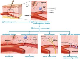
Fever
Fever is a body temperature above the normal range (36–38°C), usually in response to pyrogens released by damaged cells or bacteria.
Fever enhances immune function but is regulated by the hypothalamus.
Antimicrobial Proteins
Antibodies: Produced by B cells, function in adaptive immunity.
Complement system: Plasma proteins that mediate cell lysis and enhance inflammation and phagocytosis.
Cytokines: Regulate immune cell development and activity (e.g., interferons, interleukins, tumor necrosis factor).
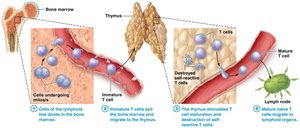
Adaptive Immunity: Cell-Mediated Immunity
T Cell Maturation and Function
T cells are formed in bone marrow and mature in the thymus, where self-reactive T cells are destroyed (self-tolerance).
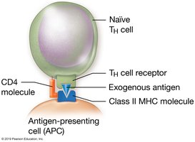
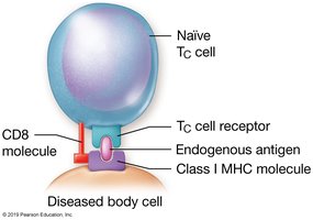
Types: Helper T (TH) cells (CD4+) interact with class II MHC molecules; Cytotoxic T (TC) cells (CD8+) interact with class I MHC molecules.
T cells respond to intracellular pathogens, cancer cells, and foreign cells (e.g., transplants).
Organ and Tissue Transplantation
Types of grafts: autografts, isografts, allografts, xenografts.
Allografts and xenografts may be rejected due to immune recognition of foreign antigens (especially MHC molecules).
Immunosuppressive therapy is used to prevent rejection.
Adaptive Immunity: Antibody-Mediated Immunity
B Cell Function and Antibody Production
B cells recognize specific antigens and differentiate into plasma cells that secrete antibodies (immunoglobulins).
Antibody-mediated immunity has three phases: antigen recognition, antibody secretion, and memory B cell formation.
Memory B cells enable a faster, stronger response upon re-exposure to the antigen (immunological memory).
Immunological Memory and Vaccination
Vaccination exposes the immune system to antigens, generating memory cells and providing long-term protection.
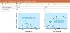
Primary immune response is slower and less robust; secondary response is faster and stronger due to memory cells.
Characteristic | Primary Immune Response | Secondary Immune Response |
|---|---|---|
Lag phase | 4–5 days | 1–3 days |
Time until antibody peak | 7–14 days | 3–5 days |
Primary antibody | IgM | IgG |
Duration of response | 14–21 days | 28 days and beyond |
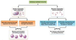
Active vs. Passive Immunity
Active immunity: Body produces its own antibodies and memory cells (via infection or vaccination); long-lasting.
Passive immunity: Preformed antibodies are transferred from another source (e.g., maternal antibodies, antibody injection); temporary, no memory cells formed.
Disorders of the Immune System
Hypersensitivity disorders: Overreaction of the immune system, causing tissue damage (e.g., allergies).
Immunodeficiency disorders: Failure of one or more components of the immune system.
Autoimmune disorders: Immune system attacks self-antigens, damaging the body's own tissues.
Summary Table: Key Structures and Functions of the Lymphatic System
Structure | Function |
|---|---|
Lymphatic vessels | Return interstitial fluid to blood, absorb dietary fats, transport immune cells |
Lymph nodes | Filter lymph, trap pathogens, house immune cells |
Spleen | Filters blood, destroys old erythrocytes, mounts immune responses |
Thymus | Site of T cell maturation |
MALT | Protects mucosal surfaces from pathogens |
