 Back
The Lymphatic System and Immunity: Structure, Function, and Clinical Relevance
Study Guide - Smart Notes
 Tailored notes based on your materials, expanded with key definitions, examples, and context.
The Lymphatic System and Immunity
Introduction to the Lymphatic and Immune Systems
The lymphatic system and immune system work together to protect the body from injury and pathogens. The lymphatic system is a network of organs and tissues that supports immune function and maintains fluid balance, while the immune system is a functional system composed of cells and proteins that defend against internal and external threats.
Immunity: Protection from injury and pathogens.
Lymphatic System: Returns interstitial fluid to the bloodstream, regulates fluid levels, absorbs dietary fats, and filters lymph and blood for pathogens.
Immune System: Defends the body using cells and proteins found in blood and tissues, including those of the lymphatic system.
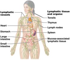
Structure and Components of the Lymphatic System
Lymphatic Vessels and Flow
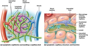
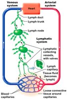
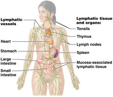
Lymphatic vessels begin as blind-ended capillaries in tissues, collecting interstitial fluid (now called lymph) and transporting it through progressively larger vessels back to the venous circulation. This system is essential for maintaining blood volume and preventing edema.
Lymphatic Capillaries: Absorb interstitial fluid and dietary fats (via lacteals in the small intestine).
Lymphatic Collecting Vessels: Transport lymph through lymph nodes for filtration.
Lymph Trunks and Ducts: Drain lymph into the venous system.
Lymphoid Cells and Tissues
Lymphoid cells are specialized for immune defense and are housed in lymphoid tissues, which provide sites for cell proliferation and immune surveillance.
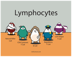
Lymphocytes: B cells, T cells, and Natural Killer (NK) cells.
Phagocytes: Macrophages and dendritic cells, which engulf pathogens and present antigens.
Reticular Cells: Produce reticular fibers that support lymphoid tissues.
Lymphoid Tissue: Loose reticular connective tissue found in organs and MALT (Mucosa-Associated Lymphatic Tissue).
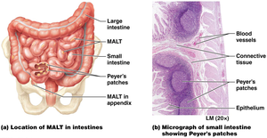
Lymphatic Organs and MALT
Lymphatic organs filter lymph and blood, and provide sites for immune cell maturation and activation. MALT protects mucosal surfaces throughout the body.
Tonsils: Trap bacteria and debris in the pharynx.
Peyer’s Patches: Defend against bacteria in the small intestine.
Appendix: Protects against bacteria in the large intestine.
Lymph Nodes: Filter lymph before it returns to the bloodstream.
Spleen: Filters blood, removes old RBCs, and houses immune cells.
Thymus: Site of T cell maturation in children.
Immune System Overview
Innate (Nonspecific) Immunity
The innate immune system provides immediate, nonspecific defense against all pathogens. It includes surface barriers and internal cellular and chemical defenses.
First Line of Defense: Skin and mucous membranes prevent pathogen entry.
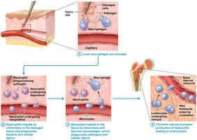
Second Line of Defense: Phagocytes, NK cells, inflammation, fever, and antimicrobial proteins respond to invaders that breach barriers.

Cells and Proteins of Innate Immunity
Phagocytes: Neutrophils and macrophages engulf and destroy pathogens.
Natural Killer (NK) Cells: Destroy infected or abnormal cells.
Dendritic Cells: Phagocytes that activate T cells.
Complement Proteins: Produced by the liver, these proteins enhance immune responses and can directly lyse pathogens.
Interferons: Proteins that inhibit viral replication.
Inflammation and Fever
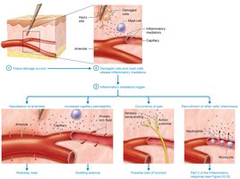
Inflammation is a tissue response to injury or infection, characterized by redness, heat, swelling, and pain. Fever is a systemic response that raises body temperature to inhibit pathogens and enhance immune activity.
Inflammatory Mediators: Histamines, cytokines, prostaglandins, and complement trigger vasodilation, increased permeability, and chemotaxis of immune cells.
Fever: Pyrogens released by leukocytes reset the hypothalamic thermostat above 38°C (99°F).
Adaptive (Specific) Immunity
Overview and Types
The adaptive immune system targets specific antigens and develops immunological memory. It is slower to respond but provides long-lasting protection.
Cell-Mediated Immunity: T cells attack infected or abnormal cells directly or via chemical signals.
Antibody-Mediated (Humoral) Immunity: B cells produce antibodies that neutralize or mark pathogens for destruction.
Lymphocyte Maturation and Antigen Recognition
T Cells: Mature in the thymus; responsible for cell-mediated immunity.
B Cells: Mature in bone marrow; responsible for antibody-mediated immunity.
Antigen: Any substance that provokes an immune response.
Antibody: Protein produced by B cells that binds specific antigens.
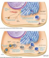
MHC Molecules: Present antigens to T cells; MHC I on most cells, MHC II on antigen-presenting cells (APCs).
Cell-Mediated Immunity
T cells require antigen presentation by MHC molecules to become activated and differentiate into functional subtypes:
Cytotoxic T Cells (CD8+): Kill infected or abnormal cells.
Helper T Cells (CD4+): Activate B cells and other immune cells.
Regulatory (Suppressor) T Cells: Suppress immune responses to prevent autoimmunity.
Memory T Cells: Provide long-term immunity.
Antibody-Mediated Immunity
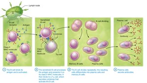
B cells are activated by antigen exposure and helper T cell signals, leading to clonal selection and differentiation into plasma cells (which secrete antibodies) and memory B cells.
Antibodies and Their Effects
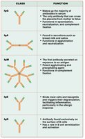
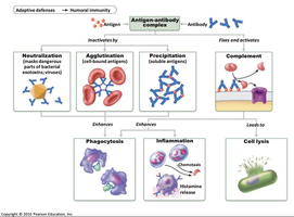
Antibodies (immunoglobulins) neutralize pathogens, promote phagocytosis, and activate complement. There are several classes of antibodies, each with specific functions.
Primary and Secondary Immune Responses
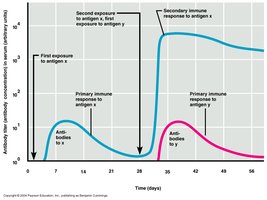
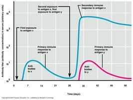
The primary response occurs upon first exposure to an antigen and is slower and weaker. The secondary response is faster and stronger due to memory cell activation.
Active and Passive Immunity
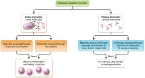
Immunity can be acquired actively (through infection or vaccination) or passively (through transfer of antibodies).
Active Immunity: Body produces its own antibodies; long-lasting.
Passive Immunity: Antibodies are received from another source; temporary protection.
Disorders of the Immune System
Allergies and Autoimmunity
Allergies: Hypersensitivity reactions to harmless substances (allergens), leading to inflammation, asthma, or anaphylactic shock.
Autoimmunity: Immune system attacks self-antigens due to loss of self-tolerance.
