 Back
BackThe Lymphatic System and Immunity: Structure, Function, and Defense Mechanisms
Study Guide - Smart Notes
 Tailored notes based on your materials, expanded with key definitions, examples, and context.
The Lymphatic System
Overview and Functions
The lymphatic system is a network of vessels and organs that plays a crucial role in maintaining fluid balance, defending the body against infection, and transporting dietary fats. It returns excess interstitial fluid to the bloodstream, filters pathogens, and supports immune responses.
Fluid Transport: Collects excess tissue fluid and returns it to the circulatory system.
Immune Defense: Filters lymph and houses immune cells that respond to pathogens.
Fat Absorption: Transports absorbed fats from the digestive tract to the bloodstream.
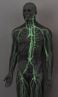
Lymphatic Pathways
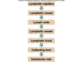
Lymphatic pathways begin with microscopic lymphatic capillaries that collect interstitial fluid, which then flows through progressively larger vessels and nodes before returning to the venous circulation.
Lymphatic Capillaries: Closed-ended tubes located in interstitial spaces; absorb tissue fluid (now called lymph).
Lymphatic Vessels: Similar to veins, with valves and three layers (endothelial lining, smooth muscle, connective tissue).
Lymph Nodes: Filter lymph and house immune cells.
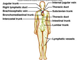
Lymphatic Trunks: Drain lymph from large regions of the body.
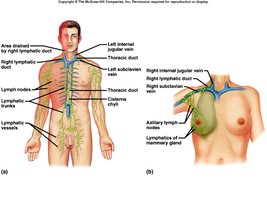
Collecting Ducts: Right lymphatic duct and thoracic duct return lymph to the subclavian veins.
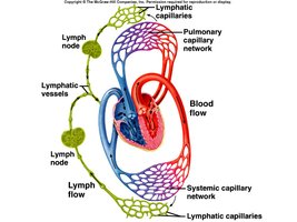
Lymphatic Capillaries and Vessels
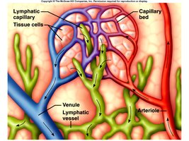
Lymphatic capillaries are highly permeable, allowing the uptake of fluid, proteins, and particulate matter. Lymphatic vessels transport lymph through nodes and eventually to the bloodstream.
Structure: Capillaries have flaplike valves; vessels have valves to prevent backflow.
Flow: Driven by skeletal muscle contraction, respiratory movements, and smooth muscle in vessel walls.
Lymphatic Trunks and Collecting Ducts
Lymphatic trunks collect lymph from vessels and drain into collecting ducts, which return lymph to the venous system.
Main Trunks: Lumbar, intestinal, intercostal, bronchomediastinal, subclavian, jugular.
Collecting Ducts: Right lymphatic duct (drains upper right body), thoracic duct (drains rest of body).
Tissue Fluid and Lymph Formation
Lymph is formed when excess tissue fluid enters lymphatic capillaries. This process prevents edema and maintains fluid balance.
Formation: Filtration from plasma exceeds reabsorption, creating tissue fluid that enters lymphatics.
Function: Prevents accumulation of excess tissue fluid (edema).
Lymph Nodes
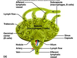
Lymph nodes are small, bean-shaped structures that filter lymph and house lymphocytes and macrophages for immune surveillance.
Structure: Surrounded by a capsule, contain nodules, sinuses, and a hilum.
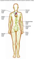
Locations: Cervical, axillary, supratrochlear, inguinal, pelvic, abdominal, thoracic regions.
Functions: Filter harmful particles, immune surveillance, lymphocyte production.
Other Lymphatic Organs
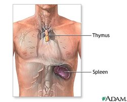
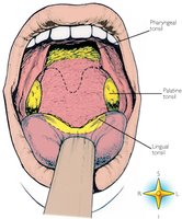
Several organs support lymphatic and immune functions, including the thymus, spleen, tonsils, and MALT (mucosa-associated lymphoid tissue).
Thymus: Site of T lymphocyte maturation; secretes thymosins.
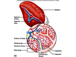
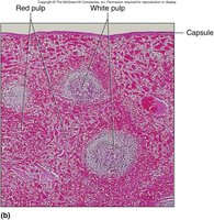
Spleen: Largest lymphatic organ; filters blood, contains white pulp (lymphocytes) and red pulp (RBCs, macrophages).
Tonsils: Lymphoid nodules in the pharynx; protect against inhaled/ingested pathogens.
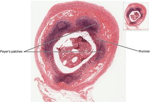
MALT: Lymphoid tissue in mucosa of GI tract, respiratory tract, etc.; includes Peyer's patches.
Immunity
Body Defenses Against Infection
The body employs both innate (nonspecific) and adaptive (specific) defenses to protect against pathogens such as bacteria and viruses.
Innate Defenses: Present at birth; general protection (e.g., skin, mucous membranes, phagocytes, inflammation, fever).
Adaptive Defenses: Develop after exposure to antigens; involve T cells and B cells for specific responses.
Innate (Nonspecific) Defenses
These defenses provide immediate, general protection against a wide range of pathogens.
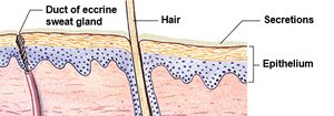
Physical Barriers: Skin, mucous membranes, tears, saliva, stomach acid.
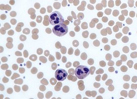
Phagocytes: Macrophages, neutrophils, eosinophils, monocytes.
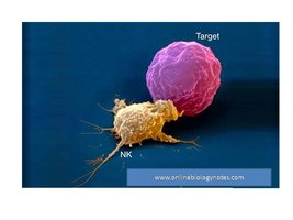
Immune Surveillance: Natural killer (NK) cells destroy abnormal cells.
Chemical Defenses: Interferons, complement system proteins.
Inflammation: Redness, swelling, heat, pain; isolates and destroys pathogens.
Fever: Raises body temperature to inhibit pathogens and speed up immune responses.
Adaptive (Specific) Defenses
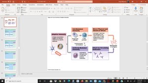
Adaptive immunity targets specific pathogens and involves memory for faster responses upon re-exposure. It is divided into cell-mediated and antibody-mediated immunity.
Cell-mediated Immunity: T cells defend against abnormal cells and pathogens inside cells.
Antibody-mediated Immunity: B cells produce antibodies that target antigens in body fluids.
Antigens and Lymphocyte Origins
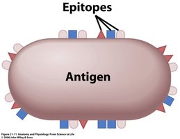
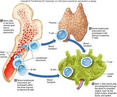
Antigens are molecules (proteins, polysaccharides, glycoproteins, glycolipids) that trigger immune responses. Lymphocytes originate from stem cells in the bone marrow and mature in either the thymus (T cells) or bone marrow (B cells).
Antigen Recognition: Helps distinguish self from non-self.
Lymphocyte Maturation: T cells mature in thymus; B cells mature in bone marrow.
T Cells and Cell-mediated Immunity
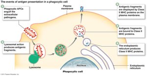
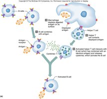
T cells must be activated by antigen presentation. Macrophages present antigens with MHC proteins, activating T cells to differentiate into helper, cytotoxic, or memory T cells.
Helper T Cells: Stimulate other immune cells.
Cytotoxic T Cells: Destroy infected or abnormal cells.
Memory T Cells: Provide long-term immunity.
B Cells and Antibody-mediated Immunity
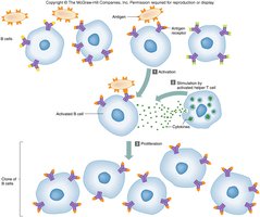
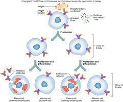
B cells are activated by helper T cell cytokines and differentiate into plasma cells (which secrete antibodies) and memory cells.
Activation: Requires antigen binding and helper T cell stimulation.
Clonal Selection: Activated B cells proliferate and differentiate.
Antibodies (Immunoglobulins)
Antibodies are Y-shaped proteins composed of four polypeptide chains. They bind specific antigens and mediate immune responses through various mechanisms.
Classes: IgG (main in blood), IgA (secretions), IgM (first produced), IgD (activates B cells), IgE (allergies).
Mechanisms: Direct attack (agglutination, precipitation, neutralization), activation of complement, inflammation.
Immune Response and Immunological Memory
The immune system mounts a primary response upon first exposure to an antigen, producing memory cells for a faster, stronger secondary response upon re-exposure.
Primary Response: Initial antibody production and memory cell formation.
Secondary Response: Rapid, robust response due to memory cells.
Types of Immunity
Immunity can be acquired naturally or artificially, and can be active (body produces its own antibodies) or passive (antibodies are received from another source).
Type | How Acquired | Example |
|---|---|---|
Naturally Acquired Active | Exposure to pathogen | Infection |
Artificially Acquired Active | Vaccination | MMR vaccine |
Naturally Acquired Passive | Mother to fetus/infant | Placenta, breast milk |
Artificially Acquired Passive | Injection of antibodies | Antivenom |
Allergic Reactions and Autoimmune Disorders
Allergic reactions are immune responses to non-harmful substances (allergens), while autoimmune disorders occur when the immune system attacks self-antigens.
Allergic Reaction: Allergen activates B cells, leading to antibody production and histamine release from mast cells.
Autoimmune Disorders: Immune system fails to recognize self, attacking body tissues (e.g., rheumatoid arthritis, type 1 diabetes).
ELISA (Enzyme-Linked Immunosorbent Assay)
ELISA is a laboratory test used to detect the presence of antigens or antibodies in a sample, commonly used for HIV screening. It relies on antigen-antibody interactions and produces a visible result.
