 Back
BackThe Lymphatic System and Lymphoid Organs: Structure, Function, and Clinical Relevance
Study Guide - Smart Notes
 Tailored notes based on your materials, expanded with key definitions, examples, and context.
The Lymphatic System: Overview
Introduction
The lymphatic system is a crucial component of the human body's defense and fluid regulation mechanisms. It returns fluids leaked from blood vessels back to the blood and provides the structural basis for the immune system by housing phagocytic cells and lymphocytes.
Lymphatic vessels: Network that drains interstitial fluid.
Lymph: Fluid within lymphatic vessels.
Lymph nodes: Cleanse lymph and provide immune surveillance.
Lymphoid organs and tissues: Include spleen, thymus, tonsils, lymph nodes, and other tissues.

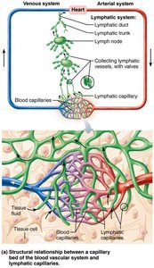
Distribution and Structure of Lymphatic Vessels
Lymphatic Capillaries and Vessels
Lymphatic vessels form a one-way system, ensuring lymph flows only toward the heart. They include lymphatic capillaries and larger collecting vessels.
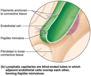
Lymphatic capillaries: Blind-ended vessels that weave between tissue cells and blood capillaries. They are absent from bones, teeth, and bone marrow, but present in the CNS meninges.
Capillaries are more permeable than blood capillaries, allowing uptake of proteins, cell debris, pathogens, and cancer cells.
Lacteals: Specialized capillaries in the intestinal mucosa that absorb digested fat and deliver fatty lymph (chyle) to the blood.
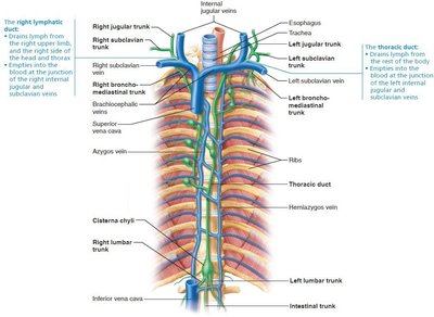
Collecting Vessels, Trunks, and Ducts
Lymph capillaries drain into larger collecting vessels, which then form lymphatic trunks and ducts.
Collecting vessels: Have thinner walls and more internal valves than veins.
Lymphatic trunks: Drain large regions of the body (lumbar, bronchomediastinal, subclavian, jugular, intestinal).
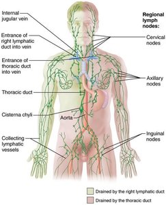
Lymphatic ducts: Right lymphatic duct drains the right upper arm and right side of head and thorax; thoracic duct drains the rest of the body.
Lymph empties into venous circulation at the junction of internal jugular and subclavian veins.
Lymph Transport
Mechanisms of Lymph Movement
The lymphatic system operates under low pressure, similar to the venous system.
Propelled by skeletal muscle contraction, thoracic pressure changes during breathing, valves preventing backflow, arterial pulsations, and smooth muscle contractions in vessel walls.
Physical activity increases lymph flow; immobilization keeps inflammatory material in place for healing.
Lymphoid Cells, Tissues, and Organs
Lymphoid Cells
Lymphoid cells include immune system cells and supporting cells.
Lymphocytes: Adaptive immune system cells, maturing into T cells (manage immune response, attack infected cells) and B cells (produce plasma cells, secrete antibodies).
Macrophages: Phagocytize foreign substances and activate T cells.
Dendritic cells: Capture antigens and deliver them to lymph nodes, activating T cells.
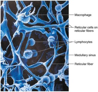
Reticular cells: Produce reticular fibers (stroma), providing scaffolding for immune cells.
Lymphoid Tissue
Lymphoid tissue houses and provides proliferation sites for lymphocytes, offering surveillance vantage points for immune cells.
Composed largely of reticular connective tissue.
Macrophages reside on reticular fibers; lymphocytes occupy spaces between fibers.
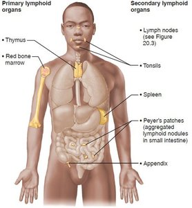
Lymphoid Organs
Diffuse lymphoid tissue: Loose arrangement of cells and fibers, found in most organs.
Lymphoid follicles (nodules): Spherical bodies with tightly packed cells and fibers, containing germinal centers of proliferating B cells.
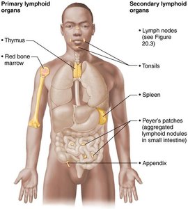
Primary lymphoid organs: Red bone marrow (B cell maturation) and thymus (T cell maturation).
Secondary lymphoid organs: Lymph nodes, spleen, MALT, and diffuse lymphoid tissues.
Lymph Nodes
Structure and Function
Lymph nodes are principal secondary lymphoid organs, found throughout the body, especially in clusters along lymphatic vessels.
Cleansing the lymph: Macrophages remove microorganisms and debris.
Immune activation: Lymphocytes become activated and mount an immune response.
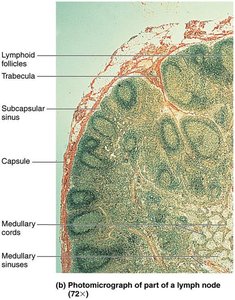
Bean-shaped, less than 2.5 cm, surrounded by a fibrous capsule.
Divided into cortex (B cell follicles, T cell transit area) and medulla (B cells, T cells, plasma cells).
Lymph sinuses contain large capillaries spanned by reticular fibers, with macrophages residing on fibers.
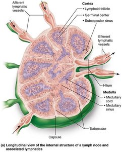
Circulation in Lymph Nodes
Lymph enters via afferent vessels, travels through sinuses, and exits via efferent vessels at the hilum.
Fewer efferent vessels cause stagnation, allowing time for immune cells to function.
Lymph passes through several nodes before returning to circulation.
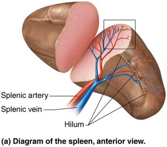
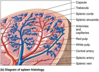
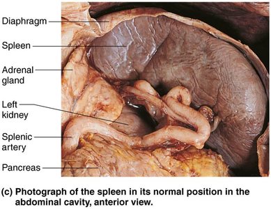
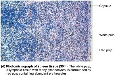
Spleen
Structure and Function
The spleen is the largest lymphoid organ, located in the left abdominal cavity.
Site of lymphocyte proliferation, immune surveillance, and response.
Cleanses blood of aged cells and platelets; stores breakdown products, platelets, and monocytes.
May produce erythrocytes in the fetus.
Histologically divided into white pulp (immune function) and red pulp (destruction of old cells and pathogens).
MALT (Mucosa-Associated Lymphoid Tissue)
Structure and Function
MALT protects mucous membranes throughout the body from pathogens.
Largest collections found in tonsils, Peyer's patches, and appendix.
Provides immune defense at entry points for pathogens.
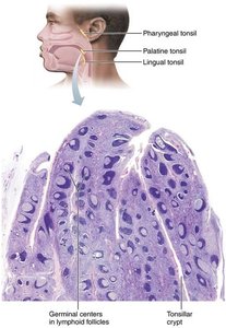
Tonsils
Form a ring of lymphatic tissue around the pharynx.
Named by location: palatine, lingual, pharyngeal (adenoids), and tubal tonsils.
Function to gather and remove pathogens in food or air.
Contain follicles with germinal centers and crypts for trapping bacteria.
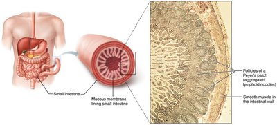
Peyer's Patches
Clusters of lymphoid follicles in the distal small intestine.
Destroy bacteria and generate memory lymphocytes.
Appendix
Contains numerous lymphoid follicles.
Functions similar to Peyer's patches: destroys bacteria and generates memory lymphocytes.
Thymus
Structure and Function
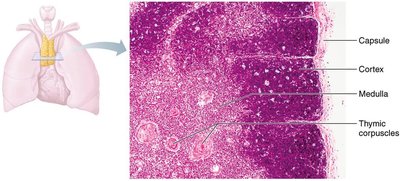
The thymus is a bilobed organ in the inferior neck and mediastinum, where T cells mature.
Most active during childhood; atrophies after adolescence but continues to produce immunocompetent cells.
Divided into cortex (rapidly dividing lymphocytes, macrophages) and medulla (fewer lymphocytes, thymic corpuscles).
Thymic corpuscles are sites for regulatory T cell development, preventing autoimmunity.
Differs from other lymphoid organs: lacks B cells, does not directly fight antigens, stroma made of epithelial cells.
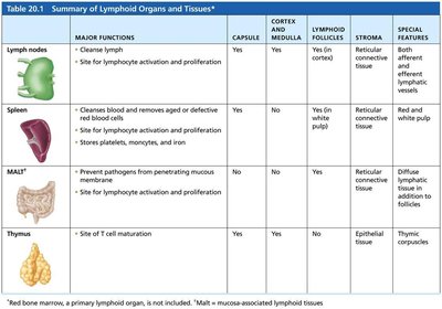
Summary Table: Lymphoid Organs and Tissues
Comparison of Major Lymphoid Organs
Organ | Major Functions | Capsule | Cortex & Medulla | Lymphoid Follicles | Stroma | Special Features |
|---|---|---|---|---|---|---|
Lymph nodes | Cleanses lymph; site for lymphocyte activation and proliferation | Yes | Yes | Yes (in cortex) | Reticular connective tissue | Both afferent and efferent lymphatic vessels |
Spleen | Cleanses blood; removes aged/defective blood cells; site for lymphocyte activation; stores platelets, monocytes, iron | Yes | Yes | Yes | Reticular connective tissue | White and red pulp |
MALT* | Prevents pathogens from penetrating mucosal surfaces; site for lymphocyte activation and proliferation | No | No | Yes | Diffuse reticular connective tissue | Collections in mucosa |
Thymus | Site of T cell maturation | Yes | Yes | No | Epithelial tissue | Thymic corpuscles |
Key Terms and Concepts
Lymph: Fluid transported by lymphatic vessels.
Antigen: Any substance recognized as foreign by the immune system.
Stroma: Supportive tissue in lymphoid organs.
White pulp: Immune function region in spleen.
Red pulp: Region in spleen for destruction of old blood cells.
Thymic corpuscles: Structures in thymus for regulatory T cell development.
Relevant Equations
Fluid Movement in Capillaries
The movement of fluid between blood capillaries and lymphatic capillaries is governed by hydrostatic and osmotic pressures:
Net filtration pressure (NFP):
Clinical Relevance
Lymphatic system dysfunction can lead to edema, impaired immunity, and spread of cancer cells.
Removal or damage to lymph nodes (e.g., during cancer treatment) can cause lymphedema.
Summary
The lymphatic system and lymphoid organs are essential for fluid balance, immune defense, and the removal of waste and pathogens. Understanding their structure and function is fundamental for students of anatomy and physiology. Additional info: Academic context and clinical relevance were added to ensure completeness and exam readiness.
