 Back
BackThe Lymphatic System and Lymphoid Organs: Structure, Function, and Clinical Aspects
Study Guide - Smart Notes
 Tailored notes based on your materials, expanded with key definitions, examples, and context.
The Lymphatic System: Overview and Functions
Introduction to the Lymphatic System
The lymphatic system is a crucial component of the circulatory and immune systems, responsible for returning interstitial fluid to the bloodstream, absorbing dietary fats, and defending the body against pathogens. It consists of a network of lymphatic vessels, lymph (the fluid), and lymphoid organs and tissues.
Returns fluids that leak from blood vessels back to the blood, maintaining fluid balance.
Absorbs fats and fat-soluble vitamins from the digestive tract and returns them to the blood.
Defends against microorganisms and disease through immune surveillance and response.
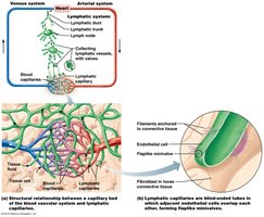
Lymphatic Vessels and Circulation
Structure and Types of Lymphatic Vessels
Lymphatic vessels form a one-way system in which lymph flows toward the heart. The system includes lymphatic capillaries, collecting lymphatic vessels, lymphatic trunks, and ducts.
Lymphatic capillaries: Highly permeable, blind-ended tubes found in most tissues except bone, teeth, bone marrow, and CNS.
Collecting lymphatic vessels: Similar to veins but with thinner walls, more valves, and more frequent anastomoses.
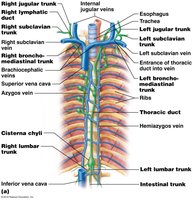
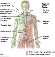
Lymphatic trunks and ducts: Major trunks drain large regions of the body and empty into two main ducts: the right lymphatic duct and the thoracic duct.
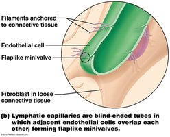
Lymphatic Capillaries and Lacteals
Lymphatic capillaries are specialized for fluid uptake and immune defense. In the intestines, lacteals absorb dietary fats, forming a milky fluid called chyle.
Flaplike minivalves: Allow interstitial fluid to enter but not exit the capillaries.
Anchoring filaments: Prevent collapse of capillaries during increased interstitial pressure.
Lymphatic Trunks and Ducts
Lymphatic trunks are named for the regions they drain (e.g., lumbar, subclavian, jugular). The right lymphatic duct drains the right upper quadrant, while the thoracic duct drains the rest of the body. The thoracic duct begins as the cisterna chyli.
Lymph Transport Mechanisms
Lymph is transported through low-pressure vessels using mechanisms similar to veins:
Milking action of skeletal muscles
Pressure changes during breathing
Valves to prevent backflow
Pulsations of nearby arteries
Contraction of smooth muscle in vessel walls
Lymphoid Cells and Tissues
Types of Lymphoid Cells
Lymphoid cells are essential for immune defense and include:
Lymphocytes: Main immune cells, divided into T cells (manage immune response, attack infected cells) and B cells (produce antibodies).
Macrophages: Phagocytize foreign substances and help activate T cells.
Dendritic cells: Capture antigens and deliver them to lymph nodes.
Reticular cells: Produce the stroma (network) that supports other cell types in lymphoid organs.
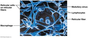
Lymphoid Tissue Structure and Function
Lymphoid tissue provides a site for lymphocyte proliferation and immune surveillance. It is primarily composed of reticular connective tissue and is found in two main forms:
Diffuse lymphoid tissue: Loose arrangement of cells and fibers, found in most organs.
Lymphoid follicles (nodules): Solid, spherical bodies with germinal centers for B cell proliferation; found in lymph nodes, Peyer's patches, and appendix.
Primary and Secondary Lymphoid Organs
Lymphoid organs are classified based on their role in lymphocyte development:
Primary lymphoid organs: Sites of lymphocyte maturation (red bone marrow for B cells, thymus for T cells).
Secondary lymphoid organs: Sites where mature lymphocytes encounter antigens (lymph nodes, spleen, tonsils, Peyer's patches, appendix).
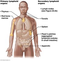
Lymph Nodes
Structure and Function of Lymph Nodes
Lymph nodes are small, bean-shaped organs that filter lymph and activate the immune system. They are clustered in regions such as the cervical, axillary, and inguinal areas.
Filtering: Macrophages remove microorganisms and debris from lymph.
Immune activation: Lymphocytes encounter antigens and mount immune responses.
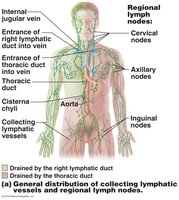
Internal Structure of Lymph Nodes
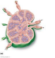
Lymph nodes have a dense capsule, trabeculae, and two main regions: cortex and medulla. The cortex contains follicles with germinal centers (B cells), while the medulla contains medullary cords (B and T cells) and sinuses (macrophages).
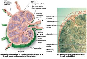
Circulation in Lymph Nodes
Lymph enters via afferent vessels, passes through sinuses, and exits via fewer efferent vessels, allowing time for immune cells to act.
Lymphoid Organs
The Spleen
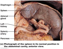
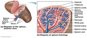
The spleen is the largest lymphoid organ, located in the left upper abdomen. It filters blood, recycles old erythrocytes, stores platelets, and is a site for immune responses.
White pulp: Site of immune function, rich in lymphocytes.
Red pulp: Site of RBC and pathogen destruction, contains splenic cords and sinusoids.
Mucosa-associated Lymphoid Tissue (MALT)
MALT consists of lymphoid tissues in mucous membranes throughout the body, providing protection at entry points for pathogens. Major MALT structures include tonsils, Peyer's patches, and the appendix.
Tonsils
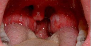
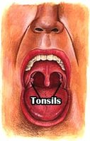
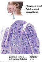
Tonsils form a ring around the pharynx and trap pathogens from food and air. They contain follicles with germinal centers and tonsillar crypts for trapping debris.
Peyer's Patches and Appendix
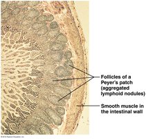
Peyer's patches are clusters of lymphoid follicles in the small intestine wall, while the appendix is a lymphoid organ attached to the large intestine. Both destroy bacteria and generate memory lymphocytes.
The Thymus
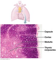
The thymus is a bilobed organ in the mediastinum, essential for T cell maturation. It is most active in childhood and atrophies with age. The thymus differs from other lymphoid organs by lacking B cells and not directly fighting antigens.
Developmental Aspects of the Lymphatic System
Lymphatic vessels and nodes develop from lymph sacs budding from veins during embryogenesis. The thymus is the first lymphoid organ to appear, followed by other organs after birth as the immune system matures.
Disorders of the Lymphatic System
Lymphedema
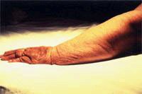
Lymphedema is chronic swelling due to lymph accumulation, often caused by obstruction or removal of lymphatic vessels.

Lymphatic Filariasis (Elephantiasis)
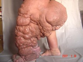
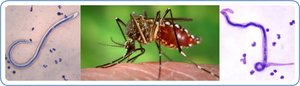
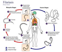
Elephantiasis is caused by parasitic roundworms that block lymphatic vessels, leading to severe swelling and thickening of the skin.
Other Disorders
Hodgkin's lymphoma: Cancer of the lymphatic system characterized by abnormal lymphocyte growth.
Castleman disease: Rare, non-cancerous lymph node disorder treated with chemotherapy.
