 Back
BackThe Lymphatic System and Lymphoid Organs: Structure, Function, and Clinical Relevance
Study Guide - Smart Notes
 Tailored notes based on your materials, expanded with key definitions, examples, and context.
The Lymphatic System: Overview and Functions
Introduction to the Lymphatic System
The lymphatic system is essential for maintaining fluid balance in the body, supporting immune function, and transporting dietary fats. It returns fluids leaked from blood vessels back to the bloodstream and provides the structural basis for the immune system.
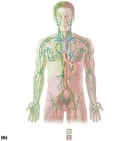
Main Components: Network of lymphatic vessels, lymph (fluid), and lymph nodes.
Lymphoid Organs and Tissues: Include the spleen, thymus, tonsils, lymph nodes, and other lymphoid tissues.
20.1 Lymphatic System Structure and Fluid Flow
Lymphatic Vessels and Circulation
Lymphatic vessels form a one-way drainage system that ensures lymph flows only toward the heart. They collect interstitial fluid and plasma proteins from tissues and return them to the blood.
Lymphatic Capillaries: Blind-ended vessels that absorb large molecules (e.g., proteins, cell debris, pathogens, cancer cells) that blood capillaries cannot.
Lacteals: Specialized lymphatic capillaries in the intestinal mucosa that absorb digested fats and deliver fatty lymph (chyle) to the blood.
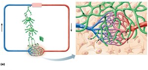
Larger Lymphatic Vessels: Lymph capillaries drain into collecting lymphatic vessels, which have thinner walls and more valves than veins. These vessels form lymphatic trunks and ducts.
Lymphatic Trunks: Drain large regions of the body and are named for the regions they serve.
Lymphatic Ducts: The right lymphatic duct drains the right upper arm and right side of the head and thorax; the thoracic duct drains the rest of the body.
Lymph Transport Mechanisms
The lymphatic system is a low-pressure system, similar to veins. Lymph is propelled by:
Milking action of skeletal muscles
Valves to prevent backflow
Contractions of smooth muscle in vessel walls
Physical activity increases lymph flow
Clinical Note: Lymphangitis is inflammation of lymphatic vessels, visible as red lines under the skin. Lymphedema is severe localized swelling due to impaired lymph return.
20.2 Lymphoid Cells, Tissues, and Organs
Lymphoid Cells
Lymphoid cells are crucial for immune defense and include both immune system cells and supporting cells.
Lymphocytes: Main cells of the adaptive immune system, divided into T cells (manage immune response, attack infected cells) and B cells (produce plasma cells that secrete antibodies).
Antibodies: Mark antigens for destruction by phagocytosis or other means.
Lymphoid Tissue
Lymphoid tissue houses lymphocytes and provides sites for immune surveillance and activation. It is mainly composed of reticular connective tissue.
Macrophages: Live on reticular fibers and phagocytose debris and pathogens.
Spaces: Allow lymphocytes to occupy and patrol the tissue.
Lymphoid Organs
Lymphoid organs are classified as primary or secondary based on their function in lymphocyte development and activation.
Primary Lymphoid Organs: Sites where T and B cells mature (red bone marrow and thymus).
Secondary Lymphoid Organs: Sites where mature lymphocytes encounter antigens (lymph nodes, tonsils, spleen, Peyer’s patches, appendix).
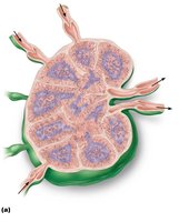
20.3 Lymph Nodes
Structure and Function of Lymph Nodes
Lymph nodes are the principal secondary lymphoid organs, distributed throughout the body. They are small, bean-shaped structures less than 2.5 cm in size.
Cleansing the Lymph: Macrophages remove and destroy microorganisms and debris, preventing unwanted substances from entering the blood.
Immune Activation: Provide sites for lymphocytes to become activated and mount immune responses.
Clinical Note: Buboes are swollen, tender lymph nodes due to overwhelming infection (e.g., bubonic plague). Cancer can also cause swollen, usually painless lymph nodes due to metastasis.
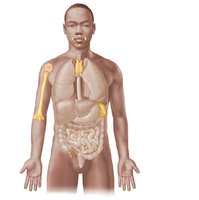
20.4 The Spleen
Structure and Functions of the Spleen
The spleen is the largest lymphoid organ, located in the left upper abdomen. It is highly vascular and serves several important functions:
Site of lymphocyte proliferation and immune surveillance
Cleanses blood by removing aged blood cells and platelets
Stores breakdown products of RBCs and platelets
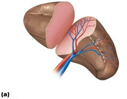
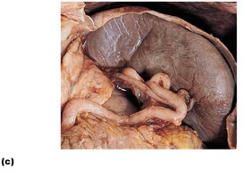
Clinical Note: The spleen has a thin capsule and is prone to rupture from trauma. Splenectomy (removal of the spleen) is less common now, as the organ can often repair itself. The liver and bone marrow can compensate for lost splenic function.
20.5 MALT (Mucosa-Associated Lymphoid Tissue)
Overview of MALT
MALT consists of lymphoid tissues in mucous membranes throughout the body, protecting against pathogens entering via mucosal surfaces. Major collections include:
Tonsils: Located in the pharynx; gather and remove pathogens in food or air.
Peyer’s Patches: Clusters of lymphoid follicles in the small intestine; destroy bacteria and generate memory lymphocytes.
Appendix: Contains many lymphoid follicles; helps destroy bacteria and generate memory lymphocytes.
Tonsils
Palatine Tonsils: Posterior end of oral cavity; largest and most often infected.
Lingual Tonsil: Base of tongue.
Pharyngeal Tonsil (Adenoids): Posterior wall of nasopharynx.
Tubal Tonsils: Surround openings of auditory tubes into pharynx.
Tonsils contain follicles with germinal centers and are not fully encapsulated. Tonsillar crypts trap pathogens, allowing immune cells to build memory.
Peyer’s Patches and Appendix
Peyer’s Patches: Located in the distal small intestine; structurally similar to tonsils.
Appendix: Offshoot of the large intestine; contains many lymphoid follicles and helps prevent bacterial breach of the intestinal wall.
20.6 Thymus
Structure and Function of the Thymus
The thymus is a bi-lobed lymphoid organ in the inferior neck, extending into the mediastinum. It is the site of T cell maturation and is most active during childhood, gradually atrophying after adolescence.
Additional info: The lymphatic system is closely integrated with the cardiovascular and immune systems, playing a vital role in fluid balance, fat absorption, and defense against pathogens and cancer.
