 Back
BackThe Lymphatic System and Lymphoid Organs and Tissues – Study Notes
Study Guide - Smart Notes
 Tailored notes based on your materials, expanded with key definitions, examples, and context.
The Lymphatic System and Lymphoid Organs and Tissues
Overview of the Lymphatic System
The lymphatic system is a network of vessels, nodes, and organs that returns fluids leaked from blood vessels back to the bloodstream and provides the structural basis for the immune system. It plays a critical role in fluid balance, fat absorption, and defense against pathogens.
Lymphatic vessels collect excess interstitial fluid (lymph) and return it to the blood.
Lymph nodes filter lymph and house immune cells.
Lymphoid organs and tissues include the spleen, thymus, tonsils, lymph nodes, and other tissues that support immune function.
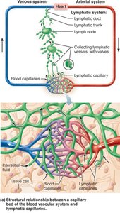
Components of the Lymphatic System
Lymph: The fluid transported by lymphatic vessels, derived from interstitial fluid.
Lymphatic vessels: Begin as blind-ended capillaries in tissues, merge into larger vessels, trunks, and ducts.
Lymph nodes: Small, bean-shaped structures that filter lymph and activate immune responses.
20.1 Lymphatic Vessels, Lymph, and Lymph Nodes
Formation and Function of Lymphatic Vessels
Hydrostatic and colloid osmotic pressures at blood capillaries force fluid out and reabsorb most of it. The remaining fluid (up to 3 L daily) becomes interstitial fluid, which is collected by lymphatic vessels.
Lymphatic vessels return protein-rich interstitial fluid to the blood.
They also transport pathogens to lymph nodes and absorbed fats from the intestine to the blood.
Structure and Distribution of Lymphatic Capillaries
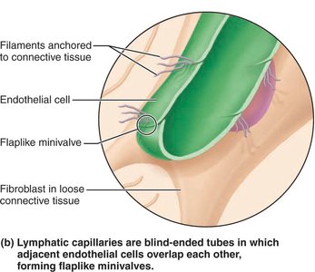
Lymphatic capillaries are highly permeable, blind-ended vessels that weave between tissue cells and blood capillaries. They are absent from bones, teeth, and most of the central nervous system (except for the glymphatic system in the brain).
Endothelial cells overlap to form one-way minivalves, allowing fluid entry but preventing backflow.
Collagen filaments anchor capillaries to surrounding tissue, so increased interstitial fluid volume opens the minivalves.
In the small intestine, specialized lymphatic capillaries called lacteals absorb digested fats, forming fatty lymph called chyle.
Larger Lymphatic Vessels, Trunks, and Ducts
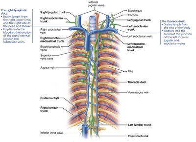
Lymphatic capillaries drain into larger collecting vessels, which have the same three tunics as veins but with thinner walls and more valves. These vessels unite to form nine major lymphatic trunks, which drain into two large ducts:
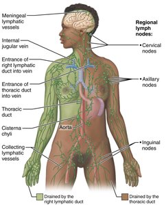
Right lymphatic duct: Drains the right upper limb, right side of head and thorax.
Thoracic duct: Drains the rest of the body; often begins as the cisterna chyli.
Distribution of Lymphatic Vessels in the Body
Clinical Correlates
Lymphangitis: Inflammation of lymphatic vessels, often visible as red streaks.
Lymphedema: Severe localized edema due to insufficient lymph return, often caused by tumors or surgical removal of lymphatics.
Lymph Transport Mechanisms
Lymphatic vessels are low-pressure conduits. Lymph flow is maintained by:
Milking action of skeletal muscles
Pressure changes during breathing
Pulsations of nearby arteries
Rhythmic contractions of smooth muscle in vessel walls
Physical activity increases lymph flow; immobilization restricts it.
20.2 Lymphoid Cells and Tissues
Lymphoid Cells
Lymphoid tissues contain immune system cells and supporting cells:
Lymphocytes: Main warriors of the immune system. Two types:
T lymphocytes (T cells): Manage immune response, attack infected cells.
B lymphocytes (B cells): Produce plasma cells that secrete antibodies.
Macrophages: Phagocytize foreign substances and activate T cells.
Dendritic cells: Capture antigens and deliver them to lymph nodes.
Reticular cells: Produce reticular fiber stroma that supports other cell types.
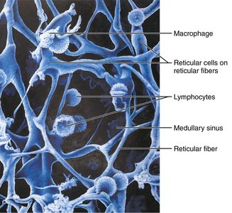
Lymphoid Tissue Structure and Types
Lymphoid tissue is primarily loose reticular connective tissue and serves as a site for lymphocyte activation and proliferation.
Diffuse lymphoid tissue: Loosely arranged, found in most organs.
Lymphoid follicles (nodules): Solid, spherical bodies with tightly packed cells, often with germinal centers for B cell proliferation.
Aggregations found in Peyer’s patches (small intestine) and appendix.
Primary and Secondary Lymphoid Organs
Primary lymphoid organs: Sites of lymphocyte maturation (red bone marrow for B cells, thymus for T cells).
Secondary lymphoid organs: Sites where mature lymphocytes encounter antigens (lymph nodes, spleen, tonsils, Peyer’s patches, appendix).
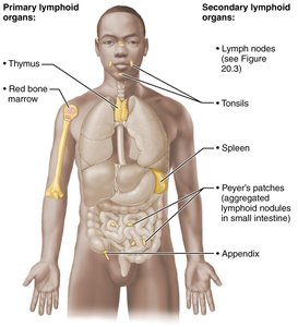
20.3 Lymph Nodes
Structure and Function of Lymph Nodes
Lymph nodes are small, bean-shaped organs clustered along lymphatic vessels, especially in the inguinal, axillary, and cervical regions. They serve two main protective functions:
Cleansing the lymph: Macrophages remove and destroy microorganisms and debris.
Immune system activation: Lymphocytes encounter antigens and mount immune responses.
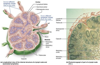
Circulation in the Lymph Nodes
Lymph enters via afferent vessels, passes through sinuses, and exits via fewer efferent vessels at the hilum.
Slow flow allows time for immune cells to act.
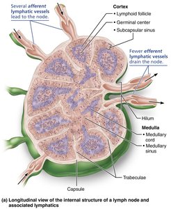
Clinical Correlates
Buboes: Infected, swollen lymph nodes (e.g., in bubonic plague).
Lymph nodes can become secondary cancer sites if metastasizing cells are trapped.
20.4 The Spleen
Structure and Functions of the Spleen
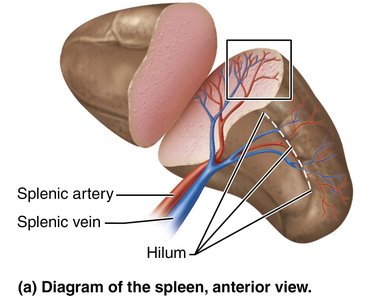
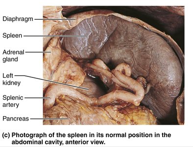
The spleen is the largest lymphoid organ, located in the left upper abdomen. It is involved in immune surveillance, blood filtration, and storage of blood components.
Removes aged red blood cells and platelets.
Stores iron, platelets, and monocytes.
Site of fetal erythrocyte production.
Histology of the Spleen
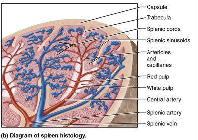
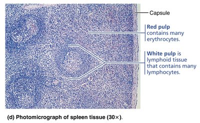
White pulp: Lymphocyte-rich regions around central arteries; site of immune function.
Red pulp: Rich in erythrocytes and macrophages; site of blood filtration and destruction of old cells.
Clinical Correlates
Splenic rupture: Can cause life-threatening hemorrhage; splenectomy may be required, but the liver and bone marrow can compensate for most functions.
20.5 MALT (Mucosa-Associated Lymphoid Tissue)
Structure and Function of MALT
MALT consists of lymphoid tissues in mucous membranes throughout the body, especially in the respiratory, digestive, and genitourinary tracts. It protects against pathogens entering through mucosal surfaces.
Major collections: Tonsils, Peyer’s patches, and appendix.
Tonsils
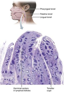
Form a ring around the pharynx; named by location (palatine, lingual, pharyngeal, tubal).
Contain follicles with germinal centers; trap and destroy pathogens in food and air.
Peyer’s Patches and Appendix
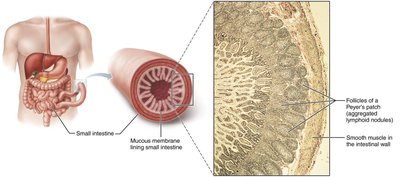
Peyer’s patches: Clusters of lymphoid follicles in the distal small intestine; prevent bacterial invasion and generate memory lymphocytes.
Appendix: Contains many lymphoid follicles; similar function to Peyer’s patches.
20.6 The Thymus
Structure and Function of the Thymus
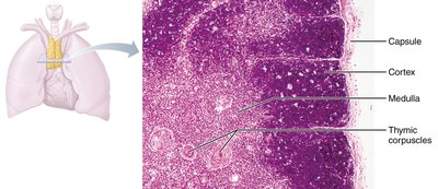
The thymus is a primary lymphoid organ located in the inferior neck and mediastinum. It is the site of T lymphocyte maturation and is most active during childhood, atrophying after puberty.
Divided into lobules with an outer cortex (rapidly dividing lymphocytes) and inner medulla (fewer lymphocytes, thymic corpuscles).
Differs from secondary lymphoid organs: lacks follicles, does not directly fight antigens, and has a blood-thymus barrier.
Stroma consists of epithelial cells, not reticular fibers.
Summary Table: Lymphoid Organs and Tissues
Organ/Tissue | Major Functions | Capsule | Cortex & Medulla | Lymphoid Follicles | Stroma | Special Features |
|---|---|---|---|---|---|---|
Lymph nodes | Cleanse lymph; site for lymphocyte activation and proliferation | Yes | Yes | Yes (in cortex) | Reticular connective tissue | Both afferent and efferent lymphatic vessels |
Spleen | Cleanses blood; removes aged RBCs; stores platelets, monocytes, iron | Yes | No | Yes (in white pulp) | Reticular connective tissue | Red and white pulp |
MALT | Prevents pathogen penetration; site for lymphocyte activation and proliferation | No | No | Yes | Reticular connective tissue | Diffuse lymphoid tissue in addition to follicles |
Thymus | Site of T cell maturation | Yes | Yes | No | Epithelial tissue | Thymic corpuscles |
Developmental Aspects of the Lymphatic System
Lymphatic vessels and main clusters of lymph nodes develop by the fifth week of embryonic life, arising from veins.
Most lymphoid organs develop from mesodermal cells, except the thymus (from endoderm).
The thymus is the first lymphoid organ to appear and is infiltrated by lymphocyte precursors before birth.
Other lymphoid organs are populated by lymphocytes shortly after birth.
