Back
The Lymphatic System and Lymphoid Organs: Structure, Function, and Clinical Relevance
Study Guide - Smart Notes
 Tailored notes based on your materials, expanded with key definitions, examples, and context.
Chapter 20: The Lymphatic System and Lymphoid Organs and Tissues
20.1 The Lymphatic System: Structure and Function
The lymphatic system is a network of vessels, nodes, and organs that returns excess tissue fluid and leaked plasma proteins to the blood, transports absorbed fats, and plays a critical role in immune defense.
Components: Lymphatic vessels, lymph (the fluid), and lymph nodes.
Primary Functions:
Return excess tissue fluid to the blood
Return leaked plasma proteins to the blood
Carry pathogens to lymph nodes for immune surveillance
Transport absorbed fats from the intestine to the blood
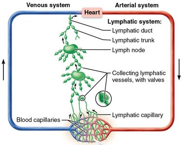
Distribution and Structure of Lymphatic Vessels
One-way system: Lymph flows only toward the heart.
Lymphatic Capillaries: Found among tissue cells and blood capillaries, especially in loose connective tissue. Absent from bones, teeth, and bone marrow.
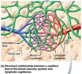
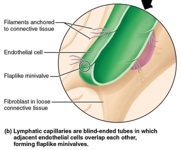
Structure of Lymphatic Capillaries
Microscopic, blind-ended tubes
Endothelial cells overlap to form flaplike mini-valves
Mini-valves open when interstitial pressure is greater than lymphatic capillary pressure, allowing fluid entry
Collagen filaments anchor endothelial cells to surrounding tissues
Function of Lymphatic Capillaries
Easily absorb excess interstitial fluid, plasma proteins, cell debris, pathogens, and cancer cells
Transport these materials to lymph nodes for immune processing
Lacteals
Specialized lymphatic capillaries in the small intestine
Transport absorbed fats (chyle) to the blood
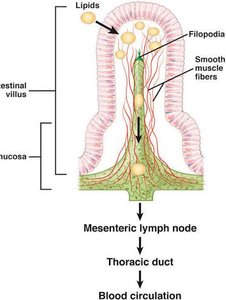
Larger Lymphatic Vessels and Flow Pathway
Lymphatic capillaries → Collecting lymphatic vessels → Lymphatic trunks → Lymph nodes → Lymphatic ducts → Venous circulation
Major lymphatic trunks: lumbar, bronchomediastinal, subclavian, jugular (paired); intestinal (single)
Right lymphatic duct drains right upper limb, right side of head and thorax; thoracic duct drains the rest of the body
Cisterna chyli: enlarged sac at the beginning of the thoracic duct, receives lymph from lumbar and intestinal trunks
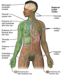
Lymph Transport Mechanisms
No central pump; relies on skeletal muscle contractions, pressure changes during breathing, one-way valves, pulsations of nearby arteries, and rhythmic contraction of smooth muscle in vessel walls
20.2 Lymphoid Cells and Tissues
Lymphoid cells and tissues are distributed throughout the body, forming the basis of immune defense.
Lymphocytes: Main warriors of the immune system; include T cells (cell-mediated immunity) and B cells (produce antibodies).
Macrophages: Phagocytize foreign substances and help activate T cells.
Dendritic cells: Capture antigens and bring them to lymph nodes.
Reticular cells: Produce reticular fiber stroma, forming the supporting network in lymphoid organs.
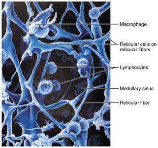
Lymphoid Tissue Structure and Function
Composed of reticular connective tissue
Houses lymphocytes and provides a site for their activation and proliferation
Continuous circulation allows rapid immune response
Types of Lymphoid Tissue
Diffuse lymphoid tissue: Loose arrangement, found in virtually every organ
Lymphoid follicles (nodules): Solid, spherical bodies with tightly packed lymphoid cells, especially B cells; found in lymph nodes, Peyer's patches, and appendix
Lymphoid Organs
Primary lymphoid organs: Sites where lymphocytes mature (red bone marrow for B cells, thymus for T cells)
Secondary lymphoid organs: Sites where mature lymphocytes encounter antigens (lymph nodes, spleen, tonsils, Peyer's patches, appendix)
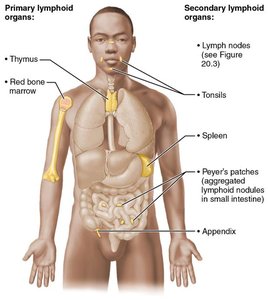
20.3 Lymph Nodes: Structure and Function
Lymph nodes are the principal secondary lymphoid organs, filtering lymph and providing sites for immune activation.
Hundreds of lymph nodes are clustered along lymphatic vessels, especially where collecting vessels converge to form trunks
Structure: Surrounded by a dense fibrous capsule; trabeculae divide the node into compartments
Cortex: Contains lymphoid follicles with dividing B cells, T cells in transit, and dendritic cells
Medulla: Contains medullary cords with B and T lymphocytes
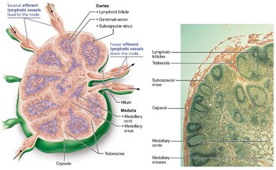
Functions of Lymph Nodes
Cleansing the lymph: Macrophages remove and destroy microorganisms and debris
Immune system activation: Lymphocytes encounter antigens and initiate immune responses
Circulation Through a Lymph Node
Lymph enters via afferent vessels → subcapsular sinus → cortical sinuses → medullary sinuses → exits at hilum via efferent vessels
Fewer efferent than afferent vessels slows lymph flow, allowing more time for immune processing
20.4 The Spleen: Structure and Function
The spleen is the largest lymphoid organ, filtering blood and providing immune surveillance.
Located on the left side of the abdominal cavity beneath the diaphragm
Functions:
Lymphocyte proliferation
Removal of bloodborne pathogens, debris, and aged RBCs/platelets
Recycles breakdown products of RBCs and stores iron
Stores platelets and monocytes
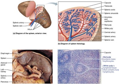
20.5 MALT: Mucosa-Associated Lymphoid Tissue
MALT is a collection of lymphoid tissues strategically located in mucous membranes to guard entryways against pathogens.
Includes tonsils, Peyer's patches, appendix, and lymphoid tissue in the mucosa of the respiratory, genitourinary, and digestive tracts
Tonsils
Form a ring of lymphoid tissue around the entrance to the pharynx
Trap and destroy bacteria and particulate matter entering in food or air
Exposure to pathogens helps produce immune cells with memory
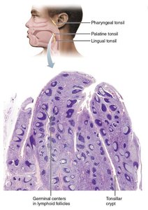
Peyer's Patches
Large clusters of lymphoid follicles in the small intestine
Prevent pathogens from breaching the intestinal wall
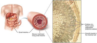
Appendix
Tubular lymphoid organ after the first part of the large intestine
Contains high concentration of lymphoid follicles
Generates memory lymphocytes for long-term immunity
20.6 The Thymus: Site of T Lymphocyte Maturation
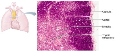
The thymus is a primary lymphoid organ where T lymphocytes mature and become immunocompetent.
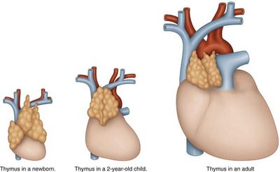
Located in the inferior neck, deep to the sternum, partially overlying the heart
Most active during childhood; atrophies after puberty but continues to produce T cells at a declining rate
Does not directly fight antigens; functions only as a maturation site for T lymphocytes
Blood-thymus barrier prevents premature activation of T cells
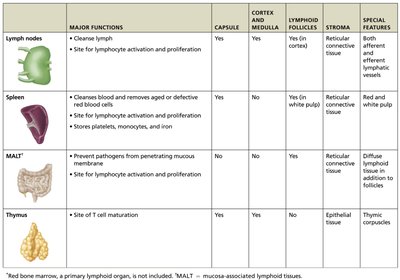
Comparative Table: Lymphoid Organs
Organ | Major Functions | Capsule | Cortex and Medulla | Lymphoid Follicles | Stroma | Special Features |
|---|---|---|---|---|---|---|
Lymph nodes | Cleanse lymph; site for lymphocyte activation and proliferation | Yes | Yes | Yes (in cortex) | Reticular connective tissue | Both afferent and efferent vessels |
Spleen | Clears blood, removes aged/defective RBCs; site for lymphocyte activation and proliferation; stores platelets, monocytes, iron | Yes | Yes (white pulp) | Yes (in white pulp) | Reticular connective tissue | Red and white pulp |
MALT | Prevents pathogens from penetrating mucous membranes; site for lymphocyte activation and proliferation | No | No | Yes | Reticular connective tissue | Diffuse lymphoid tissue in addition to follicles |
Thymus | Site of T cell maturation | Yes | Yes | No | Epithelial tissue | Thymic corpuscles |