 Back
BackThe Lymphatic System: Structure, Function, and Clinical Relevance
Study Guide - Smart Notes
 Tailored notes based on your materials, expanded with key definitions, examples, and context.
The Lymphatic System
Overview and Components
The lymphatic system is a network of cells, tissues, and organs responsible for defending the body against pathogens and maintaining fluid balance. It consists of two semi-independent parts: a network of lymphatic vessels and lymphoid organs/tissues distributed throughout the body.
Lymphatic vessels: Transport lymph, a clear fluid, from peripheral tissues to the venous system.
Lymphoid organs and tissues: Include lymph nodes, tonsils, spleen, and thymus gland.
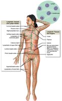
Functions of the Lymphatic System
The lymphatic system performs several essential functions:
Drainage of excess interstitial fluid: Returns fluid lost from blood capillaries back to the bloodstream.
Transport of dietary lipids and lipid-soluble vitamins: Absorbs and transports fats and vitamins (A, D, E, K) from the digestive tract.
Protection against invasion: Lymphocytes and macrophages defend against foreign cells and substances.
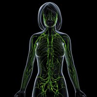
Lymphocytes and Immune Response
Types of Lymphocytes
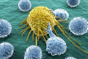
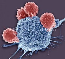
Lymphocytes are specialized white blood cells that recognize and destroy foreign substances in two main ways:
T-cells: Destroy invaders directly by releasing cytotoxic substances.
B-cells: Differentiate into plasma cells that secrete antibodies, which bind to specific foreign substances and facilitate their destruction.

Immune Response Mechanism
In specific immune responses, the lymphatic system concentrates foreign substances in certain lymphatic organs and circulates them for destruction.
Lymphatic Tissue and Nodules
Diffuse Lymphatic Tissue
Diffuse lymphatic tissue is not enclosed by a capsule and is found in small amounts in every organ, especially in the lamina propria of mucous membranes in the G.I. tract, respiratory, urinary, and reproductive systems.
Lymphoid Nodules
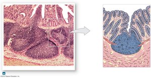
Lymphoid nodules are oval-shaped concentrations of lymphatic tissue not surrounded by a capsule, densely packed with lymphocytes. They may be solitary or found in large aggregations such as tonsils, Peyer's patches, and the appendix.
Lymphatic Vessels
Structure and Function
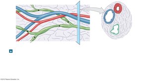
Lymphatic vessels, also called lymphatics, collect excess protein-containing interstitial fluid and return it to the bloodstream. Once interstitial fluid enters the lymphatics, it is called lymph.
Flow of Lymph
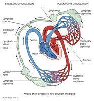
The flow of lymph follows this pathway:
Arteries (blood)
Capillaries (blood)
Interstitial spaces (interstitial fluid)
Lymphatic capillaries (lymph)
Lymphatic vessels (lymph)
Lymphatic ducts (lymph)
Subclavian veins (blood)
Lymphatic Capillaries
Lymphatic capillaries are microscopic, blind-ended vessels that weave between cells and blood capillaries. They are found throughout the body except in avascular tissues, CNS, spleen pulp, and bone marrow. These capillaries are highly permeable, allowing lymph to enter when fluid pressure is greater in the interstitial space.
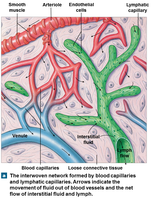
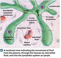
Lymphatic Vessels and Trunks
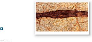
Lymphatic vessels have the same three tunics as veins but are thinner and have more internal valves. Lymphatic trunks are formed by the union of the largest collection vessels and drain large areas of the body. Major trunks include lumbar, intestinal, bronchomediastinal, subclavian, and jugular trunks.
Lymphatic Ducts
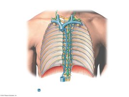
The right lymphatic duct drains lymph from the right upper arm and right side of the head and thorax, while the thoracic duct drains lymph from the rest of the body. Each duct empties lymph into the venous circulation at the junction of the internal jugular vein and subclavian vein.
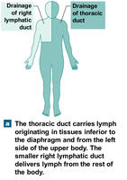
Lymph Transport
Mechanism of Lymph Movement
The lymphatic system is a pump-less system. Lymphatic vessels are low pressure, and lymph return is promoted by the milking action of skeletal muscles and pressure changes during breathing. Valves prevent backflow. Approximately 3 L of lymph enters the bloodstream every 24 hours, equal to the fluid lost to tissue spaces from the blood.
Lymphoid Organs
Primary and Secondary Lymphatic Organs
Primary lymphatic organs: Bone marrow and thymus gland; site of immunocompetent B and T cells.
Secondary lymphatic organs: Lymph nodes and spleen; most immune responses occur here.
Lymph Nodes
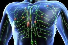
Lymph nodes are oval-shaped structures located along lymphatic vessels that filter lymph. They contain macrophages that engulf and destroy bacteria, cancer cells, and other particulate matter. Large clusters are found in the inguinal, axillary, and cervical regions.
Structure of Lymph Nodes
Stroma (framework): Capsule, trabeculae, reticular fibers, fibroblasts.
Parenchyma (inner portion): Cortex (outer) with follicles containing T-cells, macrophages, and follicular dendrite cells; medulla (inner) with lymphocytes arranged in medullary cords.
Circulation in Lymph Nodes
Lymph enters via afferent vessels, passes through sinuses in the cortex and medulla, and exits via fewer efferent vessels, allowing time for immune cells to act.
Clinical Application: Lymph Nodes and Cancer
Knowledge of lymph node location and lymph flow is crucial in cancer diagnosis and prognosis. Cancer cells may travel via the lymphatic system and form clusters of tumor cells. Cancerous lymph nodes are enlarged, firm, and non-tender, while infectious enlargement is tender.
Tonsils
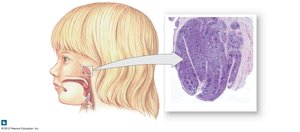
Tonsils are large aggregations of lymphatic nodules embedded in mucous membranes, forming a ring at the junction of the oral cavity and nasopharynx. They protect against inhaled or ingested foreign substances.
Pharyngeal (adenoid) tonsils: Posterior wall of nasopharynx.
Palatine tonsils: Between pharyngopalatine and glossopalatine arches.
Lingual tonsils: Base of the tongue.
Spleen
The spleen is the largest mass of lymphatic tissue, located in the abdominal cavity next to the stomach. It is surrounded by a capsule and contains white pulp (lymphatic tissue) and red pulp (venous sinuses and splenic cords).
Functions: B-cell proliferation, phagocytosis of worn-out RBCs and platelets, blood storage and release, and blood cell formation during fetal development.
Clinical Application: Ruptured Spleen
The spleen is often damaged during abdominal trauma. Rupture can cause hemorrhage and shock, requiring splenectomy. Bone marrow and liver compensate for lost spleen functions.
Thymus Gland
The thymus gland is a bi-lobed lymphatic organ in the superior mediastinum. It is covered by a connective tissue capsule, with lobules divided into cortex (site of T-cell maturation), medulla (epithelial cells and lymphocytes), and thymic corpuscles (remnants of dying cells).
Development of the Lymphatic System
The thymus is large in infants, reaches maximum size at 10-12 years, and atrophies after puberty, but continues to function in T-cell maturation. In elderly individuals, thymic tissue is mostly replaced by fat and connective tissue.
