 Back
BackThe Lymphatic System: Structure, Function, and Clinical Considerations
Study Guide - Smart Notes
 Tailored notes based on your materials, expanded with key definitions, examples, and context.
The Lymphatic System
Overview and Learning Objectives
The lymphatic system is a vital component of the body's immune and circulatory systems. It returns leaked fluid to the blood, provides anatomical support for immune defenses, and houses lymphoid organs and tissues. This section covers the structure, function, and clinical relevance of the lymphatic system.
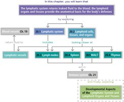
20.1 - Lymphatic System Structure
The lymphatic system consists of a network of vessels, tissues, and organs that transport lymph and support immune function.
Lymphatic Vessels: Thin-walled vessels that carry lymph, a fluid derived from interstitial fluid.
Lymphatic Capillaries: Small, blind-ended vessels that absorb excess fluid from tissues.
Lacteals: Specialized lymphatic capillaries in the intestinal mucosa that absorb dietary fats.
Large Lymphatic Vessels: Include collecting vessels, trunks, and ducts.
Lymphatic Trunks: Paired lumbar, bronchomediastinal, subclavian, jugular trunks, and a single intestinal trunk.
Lymphatic Ducts: Right lymphatic duct and thoracic duct drain lymph into the venous system.
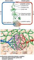
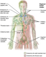
Lymph Transport
Lymph is transported through a low-pressure system similar to veins, aided by several mechanisms:
Skeletal Muscle Pump: Muscle contractions push lymph through vessels.
Thoracic Pressure: Breathing changes thoracic pressure, aiding lymph flow.
Valves: Prevent backflow of lymph.
Arterial Pulsation: Nearby arteries help propel lymph.
Smooth Muscle: Contractions in vessel walls assist movement.
20.2 - Lymphoid Cells and Tissues
Lymphoid tissues are populated by specialized cells that support immune function.
Lymphocytes: Main immune cells; include T cells (cell-mediated immunity) and B cells (antibody production).
Macrophages: Phagocytic cells that engulf pathogens and debris.
Dendritic Cells: Antigen-presenting cells that activate lymphocytes.
Reticular Cells: Support cells that form the structural framework of lymphoid tissues.
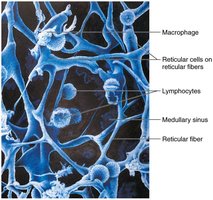
Lymphoid Organs
Primary Lymphoid Organs: Sites of lymphocyte maturation (thymus for T cells, red bone marrow for B cells).
Secondary Lymphoid Organs: Sites where mature lymphocytes encounter antigens (lymph nodes, spleen, MALT, tonsils, Peyer's patches, appendix).
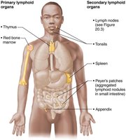
20.3 - Lymph Nodes
Lymph nodes are small, bean-shaped structures that filter lymph and house immune cells.
Regions: Cortex (outer region), medulla (inner region).
Lymph Flow: Enters via afferent vessels, passes through subcapsular and medullary sinuses, exits via efferent vessels at the hilum.
Function: Filter lymph, trap pathogens, and activate immune responses.
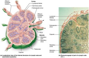
Clinical Considerations: Buboes and Lymphedema
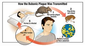
Buboes: Swollen, sometimes infected lymph nodes, often associated with bubonic plague.
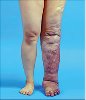
Lymphedema: Swelling due to lymphatic obstruction or dysfunction.
Phlebolymphedema: Combination of lymphedema and chronic venous insufficiency.
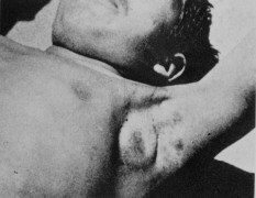
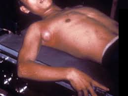
20.4 - Spleen
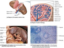
The spleen is the largest lymphoid organ, with both immune and hematological functions.
Lymphocyte Proliferation: Site for immune cell multiplication.
RBC Graveyard: Removes old and damaged red blood cells.
Stores RBC Remnants: Stores iron and other components.
Stores Platelets & Monocytes: Reservoir for blood cells.
White Pulp: Immune function (lymphocytes).
Red Pulp: RBCs and macrophages.
Fetal RBC Production: Possible site during fetal development.

Clinical Considerations: Splenomegaly
Splenomegaly: Enlargement of the spleen due to various causes (cirrhosis, hepatitis, autoimmune diseases, lymphoma, sickle cell anemia, etc.).
Pathophysiological Mechanisms: Congestive, infiltrative, immune, neoplastic, infectious (e.g., mononucleosis).

20.5 - Mucosa-Associated Lymphoid Tissue (MALT)
MALT consists of lymphoid tissues located in mucous membranes throughout the body, providing localized immune protection.
Tonsils: Remove pathogens from food and air; include palatine, lingual, pharyngeal, and tubal tonsils.
Tonsillar Crypts: Trap and destroy bacteria.
Peyer's Patches & Appendix: Destroy bacteria in the intestines and generate memory lymphocytes.

20.6 - Thymus
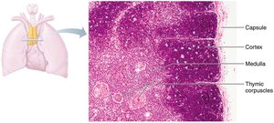
The thymus is a primary lymphoid organ essential for T lymphocyte maturation. It is most active during childhood and gradually atrophies in adulthood.
Functions: Produces immunocompetent T cells, especially regulatory T cells.
Cortex: Site of rapidly dividing lymphocytes.
Medulla: Contains thymic corpuscles where regulatory T cells develop.
Blood-Thymus Barrier: Protects developing T cells from exposure to antigens.
Summary Table: Lymphatic System Components
Component | Main Function | Location |
|---|---|---|
Lymphatic Vessels | Transport lymph | Throughout body |
Lymph Nodes | Filter lymph, immune activation | Clustered in regions (neck, axilla, groin) |
Spleen | Immune response, RBC removal | Left upper abdomen |
MALT | Local immune defense | Mucous membranes (tonsils, gut) |
Thymus | T cell maturation | Anterior mediastinum |
Key Terms and Definitions
Lymph: Fluid transported by lymphatic vessels, derived from interstitial fluid.
Lymphocyte: White blood cell involved in immune responses.
Bubo: Swollen lymph node, often due to infection.
Lymphedema: Swelling caused by lymphatic obstruction.
MALT: Mucosa-associated lymphoid tissue, provides immune protection in mucous membranes.
Splenomegaly: Enlargement of the spleen.
Equations and Formulas
There are no specific equations for the lymphatic system, but fluid movement can be described by Starling's law:
Additional info: Academic context was added to clarify the structure, function, and clinical relevance of each lymphatic system component, as well as to provide definitions and a summary table for exam preparation.
