Back
BackThe Lymphatic System: Structure, Function, and Clinical Relevance
Study Guide - Smart Notes
 Tailored notes based on your materials, expanded with key definitions, examples, and context.
The Lymphatic System: Overview
General Structure and Function
The lymphatic system is a vital component of the circulatory and immune systems, consisting of a network of lymphatic vessels and lymphoid tissues/organs. It is responsible for returning interstitial fluid and leaked plasma proteins to the blood, absorbing fats from the small intestine, and filtering/destroying foreign antigens.
Lymphatic vessels transport lymph (interstitial fluid that enters lymphatic capillaries) through a series of vessels, trunks, and ducts.
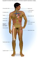
Lymphoid tissues and organs are distributed throughout the body and play key roles in immune defense.
Lymph contains proteins, microbes, and sometimes cancer cells; about 3 liters are returned to the blood daily.
Lymph movement relies on skeletal muscle and respiratory pumps.
Lymphatic Vessels and Capillaries
Lymphatic Capillaries
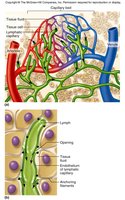
Lymphatic capillaries are similar to blood capillaries but are more permeable due to specialized endothelial minivalves. These valves function as one-way gates, allowing interstitial fluid to enter but preventing its escape, ensuring unidirectional lymph flow. During inflammation, they can absorb cell debris, pathogens, and cancer cells.
Minivalves open under increased interstitial pressure, allowing fluid entry.
Closed valves prevent backflow of lymph.
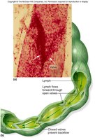
Lymphatic Collecting Vessels, Trunks, and Ducts
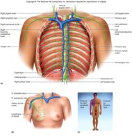
Lymphatic collecting vessels have thinner walls and more internal valves than veins, and they travel alongside arteries and veins. Nutrients are supplied by the vasa vasorum. These vessels converge to form lymphatic trunks, which then drain into two main ducts:
Right lymphatic duct: Drains the right upper arm and right side of the head and thorax.
Thoracic duct: Arises from the cisterna chyli and drains the rest of the body.
Clinical Relevance: Lymphatic Blockage and Edema
Elephantiasis and Lymphedema
Blockage of lymphatic vessels can lead to severe swelling known as lymphedema. A notable example is elephantiasis, caused by parasitic worms that clog lymph vessels, resulting in massive edema.
Symptoms include extreme swelling, most commonly in the limbs.
Chronic lymphedema can also result from surgical removal or damage to lymph nodes.


Lymphoid Tissue and Organs
Diffuse Lymphatic Tissue and Nodules
Diffuse lymphatic tissue consists of scattered reticular tissue elements found in every body organ, rich in macrophages and lymphocytes. Lymphatic nodules are solid, spherical bodies with germinal centers of maturing B cells. Examples include Peyer's patches, tonsils, and the appendix.
Mucosa-associated lymphatic tissue (MALT) is a key component of diffuse lymphatic tissue.
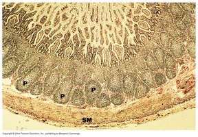
Peyer's patches are clusters of lymphoid tissue in the small intestine, especially the ileum, that destroy bacteria and prevent their entry into the intestinal wall.
Tonsils
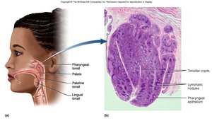
Tonsils are lymphatic nodules located in the oro/nasopharynx, filtering air and removing foreign materials. There are three types:
Pharyngeal (adenoids): Located in the nasopharynx.
Palatine: On either side of the oral cavity.
Lingual: At the root of the tongue.
Red Bone Marrow
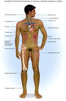
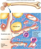
Red bone marrow is a primary lymphoid organ where B and T lymphocytes are produced, and B lymphocytes mature. In children, it is found in the medullary cavity of long bones; in adults, it is located in the axial skeleton and proximal heads of the humerus and femur. Macrophages in the marrow destroy malformed blood cells.
Thymus
The thymus is a primary lymphoid organ that secretes thymosin, promoting T lymphocyte maturation. It is largest during childhood and atrophies after adolescence. The thymus is essential for the development of the immune system.
Secondary Lymphoid Organs
Secondary lymphoid organs are sites where lymphocytes encounter pathogens and mount immune responses. These include lymph nodes and the spleen.
Lymph Nodes
Structure and Function
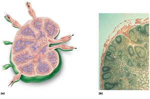
Lymph nodes are distributed throughout the body, with aggregations near the thoracic, abdominal, intestinal/mesenteric, popliteal, inguinal, axillary, and cervical regions. They swell during infection or cancer. Their two main functions are filtration and immune system activation.
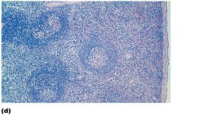
The cortex contains follicles with germinal centers rich in maturing B cells.
The deep cortex houses T cells in transit.
The medulla contains medullary cords and sinuses.
Spleen
Structure and Function
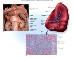
The spleen is the largest lymphoid organ. It produces B lymphocytes, cleanses the blood, stores products of RBC breakdown, salvages iron, and stores blood platelets. In fetuses, it is a site of erythrocyte production.
White pulp: Contains mostly lymphocytes and is involved in immune functions.
Red pulp: Disposes of worn-out RBCs and bloodborne pathogens.