 Back
BackThe Lymphatic System: Structure, Function, and Clinical Relevance
Study Guide - Smart Notes
 Tailored notes based on your materials, expanded with key definitions, examples, and context.
The Lymphatic System
Overview and Functions
The lymphatic system is a vital component of the circulatory and immune systems. It is responsible for draining interstitial fluid from tissues, filtering lymph to remove pathogens and debris, and returning clean lymph to the bloodstream. This system also plays a crucial role in preventing edema and facilitating immune responses.
Drains tissue fluid (interstitial fluid): Collects excess fluid from tissues and returns it to the circulatory system.
Prevents edema: By removing excess fluid, the lymphatic system prevents swelling in tissues.
Filters lymph: Lymph nodes filter out pathogens and debris before lymph is returned to the blood.
Returns lymph to circulation: Clean lymph is returned to the bloodstream at the junction of the subclavian and jugular veins.
Lymphatic Circulation
Pathway of Lymph Flow
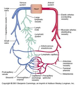
Lymphatic circulation begins with the collection of interstitial fluid by lymphatic capillaries. This fluid, now called lymph, travels through progressively larger lymphatic vessels, passing through lymph nodes for filtration, and ultimately drains into the venous system.
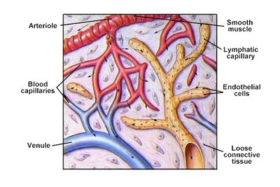
Lymphatic capillaries: Blind-ended vessels that absorb interstitial fluid from tissues.
Lymphatic vessels: Transport lymph through lymph nodes and toward larger ducts.
Lymphatic ducts: The thoracic duct and right lymphatic duct return lymph to the venous circulation.
Lymphatic Capillaries and Vessels
Structure and Function
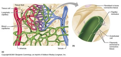
Lymphatic capillaries are highly permeable, thin-walled vessels located throughout the body. Their unique structure allows them to absorb large molecules and excess fluid from tissues. Lymphatic vessels contain valves to prevent backflow and rely on skeletal muscle contraction for lymph movement.
Endothelial cells: Overlapping cells form one-way mini-valves for fluid entry.
Anchoring filaments: Attach capillaries to surrounding tissue, preventing collapse.
Valves: Ensure unidirectional flow of lymph.
Lymph Drainage Pathways
Major Lymphatic Ducts
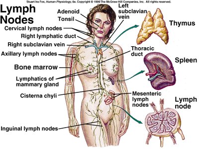
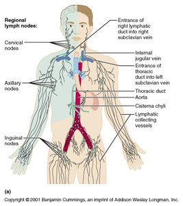
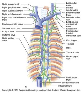
Lymph from the body is drained by two main ducts: the right lymphatic duct and the thoracic duct. These ducts empty lymph into the venous system at the junctions of the subclavian and jugular veins.
Right lymphatic duct: Drains lymph from the right upper limb, right side of the head, and right thorax.
Thoracic duct: Drains lymph from the rest of the body and is the largest lymphatic vessel.
Cisterna chyli: A dilated sac at the lower end of the thoracic duct that collects lymph from the lower limbs and abdomen.
Lymph Nodes
Structure and Function
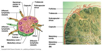
Lymph nodes are small, bean-shaped organs that filter lymph and house immune cells. They are distributed throughout the body along lymphatic vessels and are essential for immune surveillance and response.
Cortex: Contains follicles with germinal centers for B cell proliferation.
Medulla: Contains medullary cords and sinuses for lymph filtration.
Afferent and efferent vessels: Lymph enters via afferent vessels and exits via efferent vessels.
Primary and Secondary Lymphoid Organs
Thymus
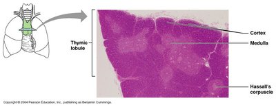
The thymus is a primary lymphoid organ located in the mediastinum. It is the site of T lymphocyte maturation and is most active during childhood.
Cortex: Densely packed with immature T cells.
Medulla: Contains mature T cells and Hassall's corpuscles.
Tonsils
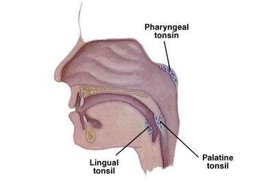
Tonsils are secondary lymphoid organs located in the pharynx. They protect against pathogens entering through the mouth and nose.
Pharyngeal tonsil (adenoids): Located in the nasopharynx.
Palatine tonsils: Located at the sides of the oropharynx.
Lingual tonsils: Located at the base of the tongue.
Spleen
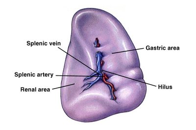
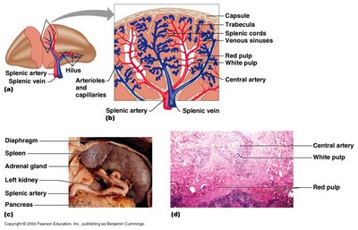
The spleen is the largest lymphoid organ and is located in the upper left abdomen. It filters blood, removes old red blood cells, and mounts immune responses to blood-borne pathogens.
White pulp: Contains lymphocytes and is involved in immune function.
Red pulp: Contains macrophages and is responsible for filtering blood and removing debris.
Hilus: Entry and exit point for splenic vessels.
Regional Lymphatics
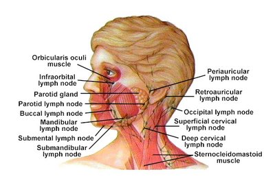
Head and Cervical Lymphatics
Lymph nodes in the head and neck filter lymph from the scalp, face, and oral cavity. They are important in detecting infections and malignancies in these regions.
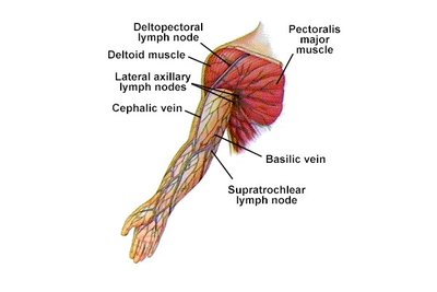
Arm and Axillary Lymphatics
Lymph from the upper limbs drains through axillary lymph nodes, which are clinically significant in breast cancer metastasis and infections of the arm.
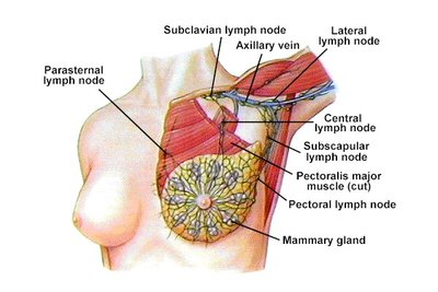
Breast Lymphatics
The lymphatic drainage of the breast is crucial for the spread of breast cancer. Lymph from the breast primarily drains into the axillary lymph nodes.
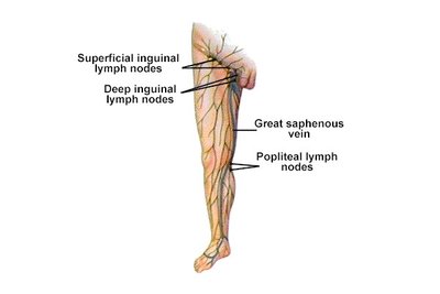
Leg and Inguinal Lymphatics
Lymph from the lower limbs drains into superficial and deep inguinal lymph nodes. These nodes are important in the detection of infections and malignancies in the lower extremities.
Colon Lymphatics
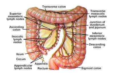
Lymphatic drainage of the colon is essential for immune surveillance and the spread of gastrointestinal cancers. Lymph from the colon drains into mesenteric lymph nodes.
Clinical Relevance: Lymphedema
Pathophysiology and Presentation
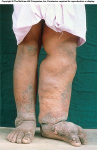
Lymphedema is the accumulation of lymph in tissues due to impaired lymphatic drainage. It results in swelling, most commonly in the limbs, and can be caused by infection, surgery, or congenital defects.
Symptoms: Swelling, heaviness, and recurrent infections in the affected limb.
Causes: Lymphatic obstruction, surgical removal of lymph nodes, or parasitic infection (e.g., filariasis).
Summary Table: Major Lymphatic Organs and Functions
Organ | Location | Main Function |
|---|---|---|
Lymph Node | Along lymphatic vessels | Filters lymph, immune surveillance |
Thymus | Mediastinum (chest) | T cell maturation |
Tonsils | Pharynx | Protects against inhaled/ingested pathogens |
Spleen | Upper left abdomen | Filters blood, immune response |
