 Back
BackThe Lymphatic System: Structure, Function, and Clinical Relevance
Study Guide - Smart Notes
 Tailored notes based on your materials, expanded with key definitions, examples, and context.
Lymphatic System Overview
Introduction to the Lymphatic System
The lymphatic system is a vital component of the circulatory and immune systems. It consists of lymphatic vessels, lymphoid organs, and tissues that work together to maintain fluid balance, filter pathogens, and support immune responses.
Lymph: The fluid transported within lymphatic vessels.
Lymphatic vessels: Network of vessels that collect and return excess interstitial fluid to the bloodstream.
Lymphoid organs and tissues: Include lymph nodes, spleen, thymus, tonsils, and mucosa-associated lymphoid tissue (MALT).
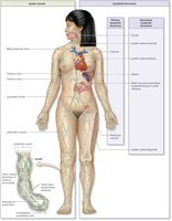
21.1 Lymph and Lymph Vessels
Functions of the Lymphatic System
The lymphatic system performs several essential functions for homeostasis and immunity:
Returns excess fluid from body tissues to the blood, maintaining blood volume.
Returns leaked proteins to the blood.
Transports and houses lymphocytes and other immune cells.
Carries pathogens to lymph nodes for filtration.
Transports absorbed fats from the intestine to the blood via specialized lymphatic capillaries called lacteals.
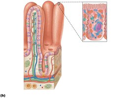
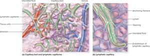
Lymphatic Capillaries and Vessels
Lymphatic capillaries are the smallest lymphatic vessels and are highly permeable due to their unique structure:
Blind-ended vessels that weave between tissue cells and blood capillaries in loose connective tissue.
Absent from bones, teeth, and bone marrow.
Endothelial cells overlap loosely to form one-way minivalves, allowing fluid to enter but not exit.
Anchored by collagen filaments to surrounding matrix, preventing collapse.
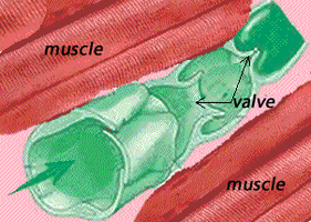
Flow of Lymph
Lymph flows in a one-way system toward the heart, propelled by:
Milking action of skeletal muscles
Pressure changes during breathing
Valves to prevent backflow
Pulsations of nearby arteries
Contraction of smooth muscle in vessel walls
Hierarchy of Lymphatic Vessels
Lymphatic capillaries drain into larger vessels in the following order:
Lymphatic capillaries
Collecting lymphatic vessels
Lymphatic trunks
Lymphatic ducts
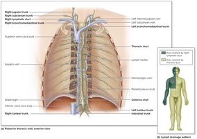
The two main lymphatic ducts are:
Right lymphatic duct: Drains right upper arm and right side of head and thorax.
Thoracic duct: Drains the rest of the body.
Both ducts empty into the venous circulation at the junction of the internal jugular and subclavian veins.
Lymphoid Cells, Tissues, and Organs
Lymphoid Cells
Lymphoid cells are crucial for immune defense and include:
Lymphocytes: Main cells of the adaptive immune system, including T cells (T lymphocytes) and B cells (B lymphocytes).
Macrophages: Phagocytize foreign substances and help activate T cells.
Dendritic cells: Capture antigens and deliver them to lymph nodes; also help activate T cells.
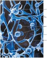
Reticular cells: Produce reticular fibers (stroma) that support other cells in lymphoid organs.
Lymphoid Tissues
Lymphoid tissues provide the structural basis for the immune system:
Diffuse lymphoid tissue: Loose arrangement of lymphoid cells and some reticular fibers, found in nearly every organ.
Lymphoid follicles (nodules): Solid, spherical bodies of tightly packed lymphoid cells and reticular fibers, often containing germinal centers for B cell proliferation.
Lymphoid Organs
Lymphoid organs are classified as primary or secondary:
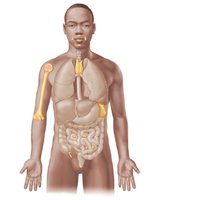
Primary lymphoid organs: Sites where T and B cells mature (red bone marrow and thymus).
Secondary lymphoid organs: Sites where mature lymphocytes first encounter antigens and become activated (lymph nodes, spleen, tonsils, Peyer's patches, appendix).
Lymph Nodes
Structure and Function
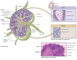
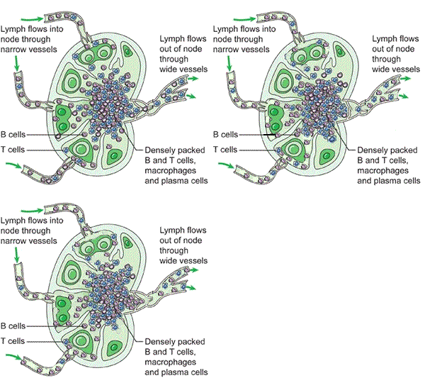
Lymph nodes are small, bean-shaped organs that filter lymph and house immune cells. They are located along lymphatic vessels, with clusters in the inguinal, axillary, and cervical regions.
Cleansing the lymph: Macrophages remove and destroy microorganisms and debris.
Immune system activation: Lymphocytes become activated and mount immune responses.
Lymph tends to stall in nodes due to fewer efferent than afferent vessels, allowing time for immune functions.
Spleen
Structure and Function
The spleen is the largest lymphoid organ and serves multiple roles:
Site of lymphocyte proliferation and immune surveillance.
Cleanses blood of aged cells and platelets; macrophages remove debris.
Stores breakdown products of RBCs (e.g., iron), blood platelets, and monocytes.
May produce erythrocytes in the fetus.
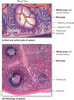
The spleen contains:
White pulp: Site of immune function, rich in lymphocytes.
Red pulp: Site where old blood cells and pathogens are destroyed, rich in RBCs and macrophages.
Mucosa-Associated Lymphoid Tissue (MALT)
Definition and Locations
MALT consists of lymphoid tissues in mucous membranes throughout the body, protecting against pathogens entering via mucosal surfaces. Major MALT structures include:
Tonsils
Peyer's patches (in the small intestine)
Appendix
Clinical Application: Edema and the Skeletal Muscle Pump
Case Study Example
A patient with swollen feet and ankles, fatigue, and shortness of breath who stands all day and is inactive may experience impaired lymphatic return. The skeletal muscle pump normally helps propel lymph through vessels by muscle contractions. Lack of movement reduces this effect, leading to fluid accumulation (edema).
Sample Questions and Answers
Lymphatic vessels collect excess fluid from interstitial spaces.
Lymphatic capillaries owe their permeability to minivalves in their walls.
Logical sequence for fluid return: Arteries, blood capillaries, lymphatic vessels, subclavian vein.
Lymph tends to stall inside lymph nodes due to fewer efferent than afferent lymphatic vessels.
Summary Table: Primary vs. Secondary Lymphoid Organs
Primary Lymphoid Organs | Secondary Lymphoid Organs |
|---|---|
Red bone marrow | Lymph nodes |
Thymus | Spleen |
Tonsils | |
Peyer's patches | |
Appendix |
Key Terms and Definitions
Lymph: Interstitial fluid that enters lymphatic vessels.
Lymphatic capillaries: Smallest lymph vessels, highly permeable due to minivalves.
Lymph nodes: Filter lymph and house immune cells.
Spleen: Filters blood, recycles RBCs, and supports immune function.
MALT: Lymphoid tissue in mucous membranes.
