 Back
BackThe Lymphatic System: Structure, Function, and Clinical Relevance
Study Guide - Smart Notes
 Tailored notes based on your materials, expanded with key definitions, examples, and context.
The Lymphatic System
Overview and Functions
The lymphatic system is a vital component of the circulatory and immune systems. It returns fluids leaked from blood vessels back to the blood, helps maintain fluid balance, and provides the structural basis for immune defense by housing phagocytic cells and lymphocytes.
Main Components:
Network of lymphatic vessels (lymphatics)
Lymph (fluid in vessels)
Lymph nodes (cleanse lymph)
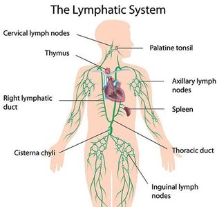
Lymphoid Organs and Tissues: Spleen, thymus, tonsils, lymph nodes, and other lymphoid tissues provide the structural basis for the immune system.
Lymphatic System Functions
Returns interstitial fluid and leaked plasma proteins to the blood via lymphatic vessels.
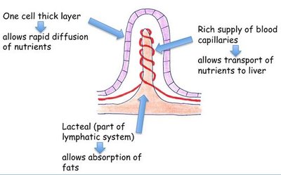
Carries absorbed fats from the digestive tract to the blood (via lacteals).
Provides a route for immune cell trafficking and surveillance.
Lymphatic Vessels and Flow
Structure and Flow of Lymph
Lymphatic vessels form a one-way system in which lymph flows only toward the heart. The system includes lymphatic capillaries, collecting vessels, trunks, and ducts.
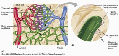
Lymphatic Capillaries: Blind-ended vessels that weave between tissue cells and blood capillaries. Absent from bones, teeth, bone marrow, and the CNS.
Collecting Lymphatic Vessels: Have thinner walls and more internal valves than veins; anastomose more frequently.
Lymphatic Trunks and Ducts: Trunks drain large areas of the body and empty into one of two ducts: the right lymphatic duct (drains right upper arm and right side of head and thorax) and the thoracic duct (drains the rest of the body).
Mechanisms of Lymph Transport
Lymph is propelled by:
Milking action of skeletal muscle
Pressure changes during breathing
Valves to prevent backflow
Pulsations of nearby arteries
Contractions of smooth muscle in lymphatic vessel walls
Physical activity increases lymph flow; immobilization slows it, aiding local immune responses.
Lymphatic Capillaries and Permeability
Specialized Structure
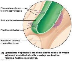
Lymphatic capillaries are highly permeable due to two specialized structures:
Endothelial cells overlap loosely to form one-way minivalves.
Minivalves open in response to increased interstitial fluid volume and close when it decreases.
Lacteals
Lacteals are specialized lymphatic capillaries in the intestinal mucosa that absorb digested fats and deliver fatty lymph (chyle) to the blood.
Lymphoid Cells and Tissues
Lymphoid Cells
Lymphocytes: Main cells of the adaptive immune system; include T cells (cell-mediated immunity) and B cells (antibody-mediated immunity).
Other Cells: Macrophages (phagocytosis), dendritic cells (antigen presentation), and reticular cells (form the stroma of lymphoid tissue).
Lymphoid Tissue Types
Diffuse Lymphoid Tissue: Loose arrangement of lymphoid cells and reticular fibers, found in nearly every organ.
Lymphoid Follicles (Nodules): Solid, spherical bodies with tightly packed lymphoid cells and reticular fibers; contain germinal centers for B cell proliferation.
Lymphoid Organs
Primary vs. Secondary Lymphoid Organs
Primary Lymphoid Organs: Sites where T and B cells mature (red bone marrow and thymus).
Secondary Lymphoid Organs: Sites where mature lymphocytes first encounter antigens and become activated (lymph nodes, spleen, MALT, tonsils, Peyer’s patches, appendix).
Lymph Nodes
Lymph nodes are the principal secondary lymphoid organs, found throughout the body, often in clusters in the inguinal, axillary, and cervical regions.
Functions:
Cleansing the lymph (macrophages remove and destroy microorganisms and debris)
Immune system activation (site for lymphocyte activation and antigen attack)
Structure: Bean-shaped, with an external fibrous capsule, trabeculae, cortex (germinal centers for B cells, T cells, dendritic cells), and medulla (sinuses, lymphocytes, macrophages).
Spleen
The spleen is the largest lymphoid organ, located in the left abdominal cavity. It is involved in lymphocyte proliferation, immune surveillance, and blood cleansing.
Removes aged blood cells and platelets; stores breakdown products, platelets, and monocytes.
Has white pulp (immune functions) and red pulp (RBC and pathogen destruction).
MALT (Mucosa-Associated Lymphoid Tissue)
MALT protects mucous membranes throughout the body and includes the tonsils, Peyer’s patches, and appendix.
Tonsils: Form a ring around the pharynx; gather and remove pathogens in food or air. Not fully encapsulated; have crypts that trap pathogens.
Peyer’s Patches: Clusters of lymphoid follicles in the small intestine wall; destroy bacteria and generate memory lymphocytes.
Appendix: Offshoot of the large intestine; contains many lymphoid follicles, with similar functions to Peyer’s patches.
Thymus
The thymus is a bilobed organ in the neck where T cells mature. It is most active in childhood and atrophies with age but continues to produce immunocompetent cells.
Has lobes with cortex (rapidly dividing lymphocytes, macrophages) and medulla (fewer lymphocytes, thymic corpuscles for regulatory T cell development).
Differs from other lymphoid organs: lacks B cells, does not directly fight antigens, and has a stroma of epithelial cells (not reticular fibers).
Clinical Relevance
Swollen Lymph Nodes (Buboes)
Inflamed, swollen, tender lymph nodes result from overwhelming infection; can become pus-filled (as in bubonic plague).
Lymph Nodes and Cancer
Lymph nodes can become secondary cancer sites if metastasizing cancer cells are trapped; these nodes are swollen but usually not painful.
Splenectomy
Surgical removal of the spleen may be necessary after rupture, but the spleen can often repair itself. Emergency splenectomies are now less common.
Appendicitis
Inflammation of the appendix can cause pain, nausea, and vomiting; rupture can lead to widespread infection and sepsis.
