 Back
The Lymphatic System: Structure, Function, and Clinical Significance
Study Guide - Smart Notes
 Tailored notes based on your materials, expanded with key definitions, examples, and context.
The Lymphatic System
Overview and Functions
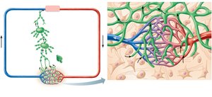
The lymphatic system is a vital component of human anatomy and physiology, responsible for maintaining fluid balance, immune defense, and the transport of lymph. It consists of lymphatic vessels, lymph, lymph nodes, and lymphoid organs.
Returns interstitial fluid and leaked plasma proteins back to the blood.
Once interstitial fluid enters lymphatics, it is called lymph.
Part of the immune system: Provides defense against pathogens and foreign substances.
Components of the Lymphatic System
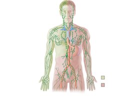
Lymphatic vessels: Network transporting lymph toward the heart.
Lymph: Fluid derived from interstitial fluid.
Lymph nodes: Principal lymphoid organs filtering lymph.
Lymphoid organs: Includes spleen, thymus, tonsils, Peyer’s patches, and appendix.
Lymphatic Vessels
Structure and Distribution
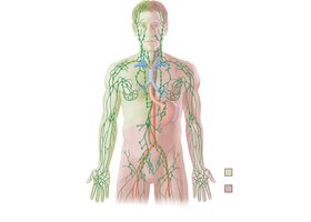
Lymphatic vessels form a one-way system, ensuring lymph flows toward the heart. There are three main types:
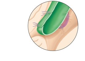
Lymphatic capillaries: Smallest, blind-ended tubes with overlapping endothelial cells forming minivalves.
Lymphatic collecting vessels: Intermediate vessels with valves.
Lymphatic trunks and ducts: Largest vessels, draining major regions of the body.
Lymphatic Ducts
Lymph is delivered into two main ducts:
Right lymphatic duct: Drains right upper arm, right side of head and thorax.
Thoracic duct: Arises from cisterna chyli, drains the rest of the body.
Each duct empties lymph into venous circulation at the junction of the internal jugular and subclavian veins.
Lymph Transport
Lymph is propelled by:
Pulsations of nearby arteries
Contractions of smooth muscle in vessel walls
Contractions of nearby skeletal muscles
Lymph transport is sporadic and slow, returning approximately 3 liters per day to the bloodstream.
Lymphoid Cells
Types and Functions
Lymphoid cells are essential for immune defense. The main types include:
Lymphocytes: Main warriors of the immune system
T cells (T lymphocytes): Manage immune response, attack and destroy foreign cells
B cells (B lymphocytes): Produce plasma cells, which secrete antibodies
Macrophages: Phagocytize foreign substances, activate T cells
Dendritic cells: Capture antigens, deliver them to lymph nodes
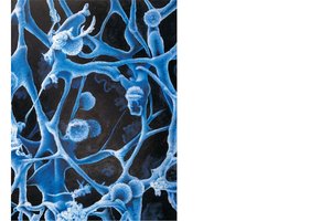
Reticular cells: Produce stroma supporting other cells in lymphoid organs
Lymphoid Tissues
Types and Functions
Lymphoid tissues provide sites for lymphocyte proliferation and immune surveillance. Two main types:
Diffuse lymphatic tissue: Scattered reticular tissue elements found in most organs
Lymphatic follicles: Solid spherical bodies with germinal centers, often part of larger lymphoid organs
Lymph Nodes
Structure and Distribution
Lymph nodes are bean-shaped organs embedded in connective tissue, clustered along lymphatic vessels, and located near body surfaces in inguinal, axillary, and cervical regions.
Functions of Lymph Nodes
Filter lymph: Macrophages destroy microorganisms and debris
Immune system activation: Lymphocytes are activated and mount an attack against antigens
Internal Structure of a Lymph Node
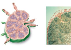
External fibrous capsule
Trabeculae: Extend inward, dividing the node into compartments
Cortex: Contains follicles with germinal centers (B cells), deep cortex houses T cells
Medulla: Medullary cords with B cells, T cells, plasma cells; lymph sinuses with macrophages
Circulation in Lymph Nodes
Lymph enters via afferent vessels
Travels through subcapsular and smaller sinuses
Exits at the hilum via efferent vessels
Fewer efferent vessels cause stagnation, allowing time for immune functions
Other Lymphoid Organs
Spleen
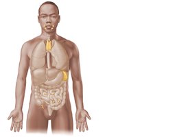
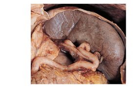
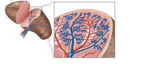
The spleen is the largest lymphoid organ, located in the upper left abdominal cavity. It is served by the splenic artery and vein.
Functions: Site of lymphocyte proliferation, immune surveillance, cleanses blood of aged cells, platelets, and debris
Stores breakdown products of RBCs (iron), blood platelets
Site of fetal erythrocyte production (ceases after birth)
Contains lymphocytes, macrophages, and erythrocytes
Structure of the Spleen
White pulp: Around central arteries, mostly lymphocytes on reticular fibers, involved in immune functions
Red pulp: Venous sinuses and splenic cords, rich in macrophages for disposal of worn-out RBCs and pathogens
Thymus
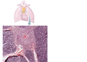
The thymus is located in the superior mediastinum and is most active during childhood.
Structure: Thymic lobes with outer cortex (densely packed lymphocytes, scattered macrophages) and inner medulla (fewer lymphocytes, Hassall’s corpuscles)
Function: T lymphocyte maturation; does not directly fight antigens
Tonsils
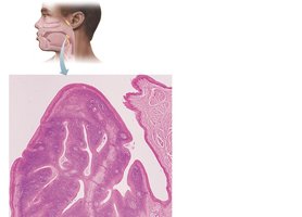
Tonsils are the simplest lymphoid organs, located in the pharyngeal region.
Contain follicles with germinal centers
Not fully encapsulated
Epithelial tissue forms tonsillar crypts, trapping and destroying bacteria
Peyer’s Patches and Appendix
Peyer’s patches: Clusters of lymphoid follicles in the distal small intestine; destroy bacteria, generate memory lymphocytes
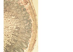
Appendix: Lymphoid follicles concentrated in the walls; similar function to Peyer’s patches
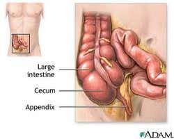
MALT (Mucosa-associated lymphatic tissue)
Includes Peyer’s patches, tonsils, appendix (digestive tract), and lymphoid nodules in bronchi (respiratory tract)
Protects passageways open to the exterior from foreign matter
Homeostatic Imbalances of the Lymphatic System
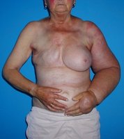
Lymphedema
Severe localized edema caused by anything preventing normal return of lymph to the blood.
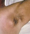
Buboes
Infected lymph nodes, commonly associated with bubonic plague. Often misnamed as "swollen glands."
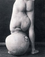
Elephantiasis
Tropical disease caused by lymphatics of lower limbs clogged with parasitic roundworms, resulting in extreme swelling.
Hodgkin’s Disease
Malignancy of lymphoid tissues
Symptoms: Swollen lymph nodes, fatigue, intermittent fevers, night sweats
Treatment: Chemotherapy and radiation
Summary Table: Major Lymphoid Organs and Their Functions
Organ | Location | Main Function |
|---|---|---|
Lymph Nodes | Clusters along lymphatic vessels | Filter lymph, immune activation |
Spleen | Upper left abdominal cavity | Immune surveillance, cleanses blood |
Thymus | Superior mediastinum | T cell maturation |
Tonsils | Pharyngeal region | Trap and destroy bacteria |
Peyer’s Patches | Distal small intestine | Destroy bacteria, memory lymphocytes |
Appendix | Large intestine | Destroy bacteria, memory lymphocytes |
Example: Lymph nodes filter lymph and activate immune responses, preventing pathogens from entering the bloodstream.
Additional info: The lymphatic system is closely integrated with the cardiovascular and immune systems, playing a critical role in homeostasis and defense against disease.
