 Back
BackThe Muscular System: Mechanisms of Skeletal Muscle Contraction and Physiology
Study Guide - Smart Notes
 Tailored notes based on your materials, expanded with key definitions, examples, and context.
The Muscular System
Overview of Skeletal Muscle Contraction
Skeletal muscle contraction is a complex process involving the interaction between nerve impulses and muscle fibers. This process is essential for voluntary movements and is regulated by a series of well-coordinated steps, from neural stimulation to the actual shortening of muscle fibers.
Nerve-Muscle Relationship: Skeletal muscles are stimulated by somatic motor neurons, which originate in the central nervous system and branch extensively to innervate muscle fibers.
Excitation: The process begins at the neuromuscular junction, where the neurotransmitter acetylcholine (ACh) is released, leading to depolarization of the muscle fiber membrane.
Contraction: The action potential travels along the sarcolemma and into T-tubules, triggering calcium release from the sarcoplasmic reticulum (SR).
Relaxation: When neural stimulation ceases, calcium is pumped back into the SR, and the muscle fiber relaxes.
Nerve-Muscle Relationship and the Neuromuscular Junction
Anatomy and Function of the Neuromuscular Junction
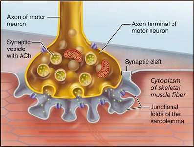
The neuromuscular junction (NMJ) is the specialized synapse where a motor neuron communicates with a skeletal muscle fiber. This junction is critical for translating neural signals into muscle action.
Axons: long, thredlike extensions of motor neurons that travel form the CNS to skeletal muscle.
Axon Terminal: The end of the motor neuron, containing synaptic vesicles filled with ACh.
The terminal and muscle fiber are seperated by the Synaptic Cleft.
Synaptic Cleft: The small gap between the axon terminal and the muscle fiber membrane.
Junctional Folds: A chemical synapse that increasae surface area and allow for neaurotransmission,
Presented as folds on the sarcolemma.
Motor End Plate: The region of the muscle fiber's membrane that interacts with the neuron.
Synaptic vesicles contain neurotransmitter acetylcholine
(ACh)
Infoldings of sarcolemma, called junctional folds, contain
millions of ACh receptors
Events at the Neuromuscular Junction
Sequence of Events Leading to Muscle Fiber Excitation
At the NMJ, the arrival of an action potential at the axon terminal triggers the release of ACh, which binds to receptors on the sarcolemma, initiating a muscle action potential.
Release of ACh: Synaptic vesicles fuse with the axon terminal membrane, releasing ACh into the synaptic cleft.
Activation of ACh Receptors: ACh binds to receptors, causing ion channels to open and sodium ions to enter the muscle fiber, generating an end plate potential (EPP).
Initiation of Action Potential: The EPP triggers an action potential that spreads across the sarcolemma.
Excitation-Contraction (E-C) Coupling
Linking Muscle Fiber Excitation to Contraction
Events that transmit AP along the sarcolemma (excitation) are coupled into sliding microfilaments (contration)
Propagation of Action Potential: The action potential travels along the sarcolemma and down T-tubules.
Calcium Release: Voltage-sensitive proteins in T-tubules stimulate the release of Ca2+ from the SR.
Initiation of Contraction: Ca2+ binds to troponin, causing tropomyosin to move and expose myosin-binding sites on actin.
Sliding Filament Model of Contraction
Mechanism of Muscle Fiber Shortening
The sliding filament model explains how muscle fibers contract by the sliding of thin (actin) filaments past thick (myosin) filaments, resulting in sarcomere shortening without a change in filament length.
Cross Bridge Formation: Myosin heads bind to actin, forming cross bridges.
Power Stroke: Myosin heads pivot, pulling actin filaments toward the center of the sarcomere.
Detachment and Recocking: ATP binds to myosin, causing it to detach from actin and recock for another cycle.
Muscle Fiber Contraction: Cross Bridge Cycling
Steps of the Cross Bridge Cycle
The cross bridge cycle is a series of molecular events that result in the sliding of actin and myosin filaments, producing muscle contraction.
Cross bridge cycling requires Calcium.
Low Ca2+ State: Tropomyosin blocks myosin-binding sites on actin; muscle is relaxed.
High Ca2+ State: Ca2+ binds to troponin, shifting tropomyosin and exposing binding sites.
When the cycle stops, the stimulation of Calcium ceases and is pumped back into the SR.
Cycle Steps:
Cross bridge formation (myosin binds to the thin actin)
Power stroke (myosin head pivots, pulling actin toward the M line)
Cross bridge detachment (ATP binds myosin, releasing actin)
Cocking of myosin head (ATP hydrolysis repositions myosin into a high energy state)
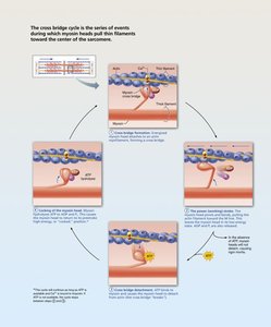
One way to think about it: 1. Grab the rope, 2. pull the rope "power stroke", 3. let go "cross bridge detachment", 4. reach forward "cock myosin head"
SUMMARY:
Key Steps in Muscle Contraction (Sliding Filament Model)
Neural Stimulus: A signal travels to the neuromuscular junction, releasing acetylcholine (ACh).
The signal is an impulse called an action potential and travels through a type of nerve cell called a motor neuron.
When the nervous system signal reaches the neuromuscular junction a chemical message is released by the motor neuron. The chemical message, a neurotransmitter called acetylcholine, binds to receptors on the outside of the muscle fiber. That starts a chemical reaction within the muscle.
Calcium Release: The action potential causes the sarcoplasmic reticulum to release calcium ions (Calcium) into the muscle cell.
Binding Site Exposure: Calcium binds to troponin, which shifts tropomyosin, exposing binding sites on the actin filament.
These are regulatory proteins that block the binding of myosin to actin. Calcium causes them to move.
Cross-Bridge Formation: Myosin heads bind to the exposed sites on actin.
a. Cross bridge formation (myosin binds to the thin actin)
b. Power stroke (myosin head pivots, pulling actin toward the M line)
Myosin releases ADP and phosphate, bending and pulling the actin filament toward the center of the sarcomere.
c. Cross bridge detachment (ATP binds myosin, releasing actin), a new ATP molecule binds to the myosin head, causing it to detach from actin.
d. Cocking of myosin head (ATP hydrolysis repositions myosin into a high energy state)
Relaxation (Detachment): A new ATP molecule binds to the myosin head, causing it to detach from actin.
Repetition: As long as calcium and ATP are present, the cycle repeats, shortening the muscle.
Clinical Homeostatic Imbalances
Disorders Affecting Neuromuscular Function
Several diseases and conditions can disrupt normal neuromuscular function, leading to muscle weakness or abnormal contraction.
Myasthenia Gravis: An autoimmune disease where antibodies destroy ACh receptors, resulting in muscle weakness and fatigue.
Characterized by drooping eyelids, difficulty swallowing and talking, and muscle weakness.
Antibodies attack the Ach receptors making it an autoimmune disease.
Rigor Mortis: After death, ATP production ceases, preventing Ca2+ reuptake and cross bridge detachment, causing muscles to stiffen until proteins degrade.
Occurs 3-4 hours after death.
The muscles begin to stiffin due to intracellular calcium levels increases without being pumped back in. Simply, mucles are in a constant state of contraction.
Whole Muscle Contraction
Principles of Muscle Tension and Load
Muscle contraction produces tension, which is the force exerted by a muscle on an object.
The load is the opposing force exerted by the object.
The force and duration of contraction depend on the frequency and intensity of stimulation.
Contraction may/may not shorten muscle
Isometric Contraction: No shortening, muscle tension increases but does not exceed load.
Isotonic Contraction: Muscle Shortens, muscle tension exceeds the load.
Isotonic contractions can be either concentric or eccentric:
Concentric contractions: muscle shortens and does work
Example: biceps contract to pick up a book
Eccentric contractions: muscle lengthens and generates force
Example: laying a book down causes biceps to lengthen while generating a force
Muscle Metabolism
ATP Production Pathways in Muscle
Muscle contraction requires ATP (energy), which can be generated through several metabolic pathways:
Anaerobic Fermentation: Glucose is converted to lactate, producing 2 ATP per glucose without oxygen.
Useful for short bursts of activity but yields little ATP, cannot support endurance activities.
Does not require oxygen.
example: sprinting
Aerobic Respiration: Glucose is fully oxidized in mitochondria, producing up to 30 ATP per glucose with oxygen.
More efficient but slower and requires oxygen.
example: lifting heavy weights
Phosphagen System: Creatine phosphate donates a phosphate to ADP to rapidly regenerate ATP
Appropriate for short, intense activity.
Fatigue and Endurance
Factors Affecting Muscle Performance
Muscle fatigue: the decline in ability to generate force, often due to prolonged activity. Endurance is influenced by metabolic and physiological factors.
Causes of Fatigue: Glycogen depletion, calcium leakage, potassium accumulation, and reduced excitability.
Endurance Factors: Myoglobin and glycogen stores, capillary density, mitochondrial number, oxygen uptake, and psychological willpower.
Types of Muscle Fibers
Classification and Functional Differences
Muscle fibers are classified based on their contraction speed and resistance to fatigue:
Slow-Twitch (Type I): Contract slowly, resist fatigue,
adapted for aerobic respiration, rich in mitochondria, myoglobin, and capillaries.
endurance based activity with oxygen
Fast-Twitch (Type II): Contract quickly, fatigue rapidly,
adapted for anaerobic respiration, rich in enzymes for glycolysis, and have a more extensive SR.
short burst activity without oxygen
Muscular Strength and Conditioning
Effects of Exercise on Muscle Tissue
Muscle strength and endurance can be improved through different types of exercise, but muscle cells do not increase in number, only in size (hypertrophy).
Resistance Training: Increases muscle size and strength but not fatigue resistance.
Endurance (Aerobic) Exercise: Improves fatigue resistance, increases glycogen, mitochondria, and capillary density, but does not significantly increase strength.
Cross-Training: Combines both types for optimal performance.
Sample Questions and Key Facts
When osteoblasts become enclosed in lacunae, they become osteocytes.
Alternating light and dark bands in skeletal muscle are called striations.
One motor neuron typically stimulates multiple muscle fibers, but each muscle fiber is innervated by only one motor neuron.
The excitatory neurotransmitter for skeletal muscle is acetylcholine.
