 Back
The Muscular System: Skeletal Muscle Tissue and Muscle Organization
Study Guide - Smart Notes
 Tailored notes based on your materials, expanded with key definitions, examples, and context.
The Muscular System: Skeletal Muscle Tissue and Muscle Organization
Introduction to Muscle Tissue
Muscle tissue is specialized for contraction and is essential for movement, posture, and various bodily functions. There are three main types of muscle tissue, each with distinct characteristics and roles in the human body:
Skeletal Muscle: Attaches to bones and is responsible for voluntary movements.
Cardiac Muscle: Found only in the heart, responsible for pumping blood through rhythmic contractions.
Smooth Muscle: Located in the walls of hollow organs (e.g., digestive tract), responsible for involuntary movements of fluids and solids.
Basic Properties of Muscle Tissue
All muscle tissues share four fundamental properties that enable their function:
Excitability: The ability to respond to stimuli.
Contractility: The ability to shorten and generate force.
Extensibility: The ability to be stretched without being damaged.
Elasticity: The ability to return to original length after contraction or extension.
Functions of Skeletal Muscles
Skeletal muscles perform several vital functions in the body:
Produce skeletal movement
Maintain posture and body position
Support soft tissues
Regulate entry and exit of materials (e.g., sphincters)
Maintain body temperature through heat production
Anatomy of Skeletal Muscles
Connective Tissue Components
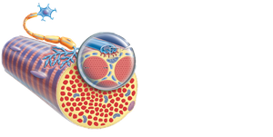
Skeletal muscles are organized and supported by three layers of connective tissue:
Epimysium: Dense connective tissue surrounding the entire muscle.
Perimysium: Divides the muscle into bundles called fascicles.
Endomysium: Surrounds individual muscle fibers (cells).
These connective tissues converge to form tendons (attach muscle to bone) or aponeuroses (broad, flat sheets connecting muscle to muscle).
Muscle Structure: From Organ to Cell
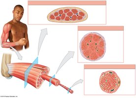
Skeletal muscle is organized hierarchically from the whole muscle down to the molecular level:
Muscle (organ): Surrounded by epimysium, contains bundles of fascicles.
Fascicle: Bundle of muscle fibers, surrounded by perimysium.
Muscle fiber (cell): Surrounded by endomysium, contains myofibrils.
Myofibril: Composed of myofilaments (actin and myosin).
Nerves and Blood Vessels
Nerves penetrate the epimysium to innervate muscle fibers, while blood vessels run parallel to nerves, supplying oxygen and nutrients. The neuromuscular junction is the site where a motor neuron communicates with a muscle fiber to initiate contraction.

Microanatomy of Skeletal Muscle Fibers
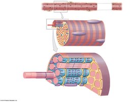
Each muscle fiber is a multinucleated cell with specialized structures:
Sarcolemma: Plasma membrane of the muscle cell.
Sarcoplasm: Cytoplasm of the muscle cell.
Transverse (T) tubules: Invaginations of the sarcolemma that transmit action potentials into the cell.
Myofibrils: Cylindrical structures responsible for contraction, composed of myofilaments (actin and myosin).
Sarcoplasmic reticulum (SR): Specialized endoplasmic reticulum that stores calcium ions, essential for muscle contraction.
Sarcomere Structure
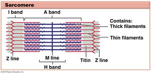
The sarcomere is the functional unit of muscle contraction, defined as the segment between two Z lines. It contains organized arrays of thick (myosin) and thin (actin) filaments, which slide past each other during contraction.
Z line: Boundary of each sarcomere.
A band: Region containing thick filaments.
I band: Region containing only thin filaments.
H band: Central region with only thick filaments.
M line: Center of the sarcomere.
Motor Units and Muscle Control
Motor Units
A motor unit consists of a single motor neuron and all the muscle fibers it innervates. The size of the motor unit determines the precision of muscle control:
Small motor units: Precise control (e.g., eye muscles).
Large motor units: Less precise, more powerful contractions (e.g., thigh muscles).
Muscle tension depends on the frequency of stimulation and the number of motor units recruited (recruitment). Muscle tone is the baseline tension in a relaxed muscle, important for posture and joint stability.
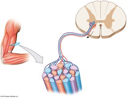
Organization of Skeletal Muscle Fibers
Muscle Fiber Arrangements
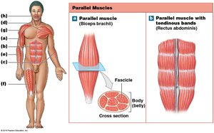
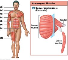
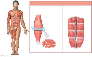
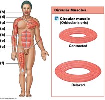
Muscles are classified by the arrangement of their fibers, which affects their function and force generation:
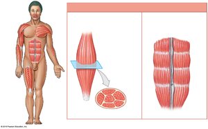
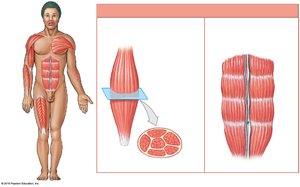
Parallel: Fibers run parallel to the long axis (e.g., biceps brachii, rectus abdominis).
Convergent: Fibers converge to a single tendon (e.g., pectoralis major).
Pennate: Fibers attach obliquely to a tendon (unipennate, bipennate, multipennate; e.g., extensor digitorum, rectus femoris, deltoid).
Circular: Fibers arranged in concentric rings (e.g., orbicularis oris).

Muscle Terminology
Origins, Insertions, and Actions
Muscle attachments and actions are described as follows:
Origin: The fixed attachment point (usually proximal).
Insertion: The movable attachment point (usually distal).
Action: The movement produced when the muscle contracts.
Muscles are grouped by their actions:
Prime mover (agonist): Main muscle responsible for a movement.
Antagonist: Opposes the action of the agonist.
Synergist: Assists the prime mover.
Fixator: Stabilizes the origin of the prime mover.
Example: In forearm flexion, the biceps brachii is the prime mover, triceps brachii is the antagonist, and muscles like the brachialis act as synergists.
Naming Muscles
Muscles are named based on various criteria:
Location (e.g., abdominis, brachii)
Shape (e.g., deltoid, trapezius)
Fiber orientation (e.g., rectus)
Number of heads (e.g., biceps, triceps)
Origin and insertion (e.g., sternocleidomastoid)
Function (e.g., extensor digitorum)
