 Back
BackThe Muscular System: Structure and Function
Study Guide - Smart Notes
 Tailored notes based on your materials, expanded with key definitions, examples, and context.
The Muscular System
Introduction to the Muscular System
The muscular system is essential for generating force and enabling movement throughout the body. Muscles are responsible for actions such as walking, breathing, pumping blood, and moving food through the digestive tract. There are three types of muscle tissue: skeletal, smooth, and cardiac muscle.
Structure of Skeletal Muscle
Connective Tissue Coverings
Skeletal muscles are composed of over 600 individual muscles, each surrounded and separated by layers of dense connective tissue called fascia. Fascia extends beyond the muscle, forming tendons that attach to bones, or broad sheets called aponeuroses that connect muscles to each other. The connective tissue layers include:
Epimysium: Surrounds the entire muscle
Perimysium: Surrounds bundles of muscle fibers called fascicles
Endomysium: Covers each individual muscle fiber
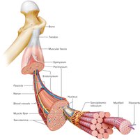
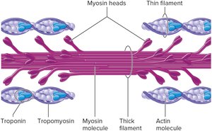
Skeletal Muscle Fibers
Each muscle fiber is a single, long, cylindrical cell that responds to stimulation by exerting a pulling force. The cell membrane is called the sarcolemma, and the cytoplasm is the sarcoplasm, which contains many mitochondria and nuclei. Within the sarcoplasm are parallel myofibrils composed of thick (myosin) and thin (actin, troponin, tropomyosin) filaments. The arrangement of these filaments produces visible bands called striations.
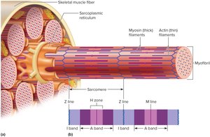
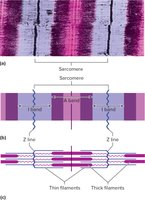
Myofibrils and Sarcomeres
Myofibrils are made up of repeating units called sarcomeres, which extend from one Z line to the next. Striations are formed by alternating light (I bands) and dark (A bands) regions:
I bands: Light bands, composed of actin filaments anchored to Z lines
A bands: Dark bands, composed of overlapping thick and thin filaments
H zone: Central region of the A band, contains only myosin filaments
M line: Center of the H zone, holds myosin filaments in place
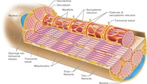
Sarcoplasm and Associated Structures
Beneath the sarcolemma lies the sarcoplasmic reticulum (SR), a network of membranous channels that stores calcium ions. The SR is closely associated with transverse (T) tubules, which are invaginations of the sarcolemma. T tubules are open to the outside of the muscle fiber and, together with the SR, play a crucial role in activating muscle contraction.
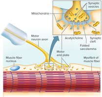
Neuromuscular Junction
Structure and Function
Skeletal muscle fibers contract only when stimulated by a motor neuron. The functional connection between a motor neuron and a muscle fiber is called a neuromuscular junction. At this synapse, the neuron releases neurotransmitters (primarily acetylcholine) that bind to receptors on the muscle fiber, triggering contraction.
Skeletal Muscle Contraction
Sliding Filament Model
Muscle contraction is based on the sliding filament model, where myosin heads bind to actin filaments, forming cross-bridges. This process pulls actin filaments toward the center of the sarcomere, shortening the muscle fiber and generating force. The contraction cycle is powered by ATP, which is hydrolyzed by the enzyme ATPase.
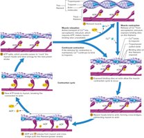
Major Events of Muscle Contraction and Relaxation
Acetylcholine is released at the neuromuscular junction
Sarcoplasmic reticulum releases calcium ions
Calcium binds to troponin, moving tropomyosin and exposing myosin binding sites on actin
Myosin heads form cross-bridges and pull actin filaments
ATP binds to myosin, breaking the cross-bridge and allowing relaxation
After death, muscles contract and become rigid (rigor mortis) due to increased calcium permeability and decreased ATP.
Energy Sources for Contraction
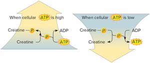
ATP and Creatine Phosphate
ATP is the immediate source of energy for muscle contraction, but it is limited and must be regenerated. Creatine phosphate serves as a rapid means to regenerate ATP from ADP and phosphate. When ATP levels are high, creatine phosphate is synthesized; when ATP levels are low, creatine phosphate donates its phosphate to ADP, forming ATP.
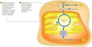
Cellular Respiration and Oxygen Supply
Muscle cells rely on cellular respiration to generate ATP. Glycolysis (anaerobic) yields 2 ATP per glucose, while aerobic respiration (in mitochondria) yields 28 ATP per glucose. Oxygen is delivered by hemoglobin and stored in muscle by myoglobin, enhancing aerobic capacity.
Oxygen Debt
During strenuous exercise, oxygen supply may be insufficient, leading to accumulation of lactic acid and the development of oxygen debt. Oxygen debt is the amount of oxygen required to convert lactate back to glucose and restore ATP and creatine phosphate levels. This process is also known as excess post-exercise oxygen consumption (EPOC).
Muscular Responses
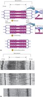
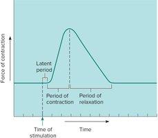
Muscle Twitch and Myogram
The response of a single muscle fiber to a single stimulus is called a twitch, which consists of contraction and relaxation phases. A myogram records these events, showing the latent period, contraction, and relaxation.
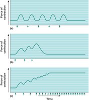
Summation and Tetanic Contraction
When a muscle fiber receives repeated stimuli, the force of contraction increases through summation. If stimuli are frequent enough, the muscle may enter partial tetany (incomplete relaxation) or complete tetanic contraction (no relaxation).
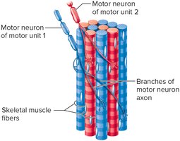
Recruitment of Motor Units
A motor unit consists of a motor neuron and the muscle fibers it controls. Increasing the number of activated motor units (recruitment) increases the strength of contraction. Maximum tension is achieved when all motor units are recruited.
Skeletal Muscle Actions
Origin, Insertion, and Movement
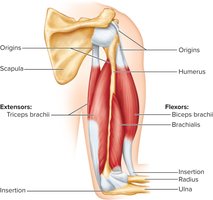
The origin of a muscle is its less movable end, while the insertion is the more movable end. Muscle contraction pulls the insertion toward the origin. Muscles may have multiple origins or insertions, as seen in the biceps brachii.
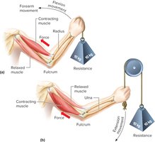
Levers and Movement
Bones and muscles interact as levers to produce movement. The parts of a lever include:
Rigid bar: bone
Fulcrum: joint
Force: muscle contraction
Resistance: object moved
Major Skeletal Muscles
Anterior and Posterior Views
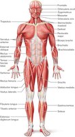
Skeletal muscles are named based on size, shape, location, action, number of attachments, and direction of fibers. Examples include pectoralis major, deltoid, extensor digitorum, biceps brachii, sternocleidomastoid, and external oblique.

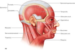
Muscles of Facial Expression
Key Muscles
Muscles of facial expression attach to bones and connective tissue of the skin, enabling a wide range of expressions. Major muscles include epicranius, orbicularis oculi, orbicularis oris, buccinator, zygomaticus, and platysma.
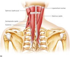
Muscles That Move the Head
Key Muscles
Paired muscles in the neck and upper back cause flexion, extension, and rotation of the head. Major muscles include sternocleidomastoid, splenius capitis, semispinalis capitis, and scalenes.
Muscles That Move the Pectoral Girdle
Key Muscles
Muscles that move the pectoral girdle include trapezius, rhomboid major, levator scapulae, serratus anterior, and pectoralis minor.
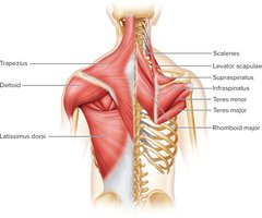

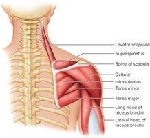
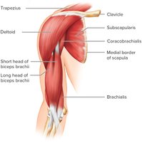
Muscles That Move the Arm
Key Muscles
Muscles connecting the arm to the pectoral girdle, ribs, and vertebral column are grouped by action: flexors (coracobrachialis, pectoralis major), extensors (teres major, latissimus dorsi), abductors (supraspinatus, deltoid), and rotators (subscapularis, infraspinatus, teres minor).
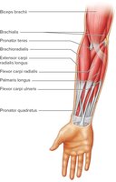
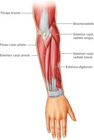
Muscles That Move the Forearm
Key Muscles
Forearm movements are accomplished by muscles arising from the humerus or pectoral girdle and connecting to the ulna and radius. Flexors include biceps brachii, brachialis, and brachioradialis; the extensor is triceps brachii; rotators include supinator, pronator teres, and pronator quadratus.
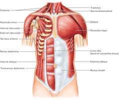
Muscles of the Abdominal Wall
Key Muscles
The abdominal wall is supported by broad, flattened muscles connecting the rib cage and vertebral column to the pelvic girdle. The four muscles are external oblique, internal oblique, transverse abdominis, and rectus abdominis. The linea alba is a band of connective tissue serving as an attachment point.
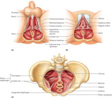
Muscles of the Pelvic Floor
Key Muscles
Two muscular sheets close off the inferior outlet of the pelvis: the pelvic diaphragm (levator ani, coccygeus) and the urogenital diaphragm (superficial transverse perineal, bulbospongiosus, ischiocavernosus).
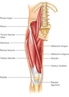
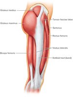
Muscles That Move the Thigh
Key Muscles
Muscles that move the thigh are attached to the femur and pelvic girdle and are classified as anterior (flexors: psoas major, iliacus), medial (adductors: adductor magnus, adductor longus, gracilis), and posterior (extensors, abductors, rotators: gluteus maximus, gluteus medius, gluteus minimus, tensor fasciae latae).
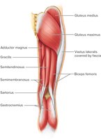
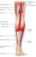
Muscles That Move the Leg
Key Muscles
Muscles connecting the tibia or fibula to the femur or pelvic girdle are grouped as flexors (hamstring group: biceps femoris, semitendinosus, semimembranosus, sartorius) and extensors (quadriceps femoris group: rectus femoris, vastus lateralis, vastus medialis, vastus intermedius).


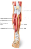
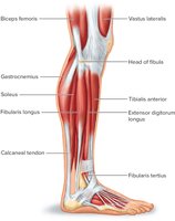
Muscles That Move the Foot
Key Muscles
Muscles that move the foot are attached from the femur, fibula, or tibia to bones of the foot. Movements include dorsiflexion (tibialis anterior, fibularis tertius, extensor digitorum longus), plantar flexion (gastrocnemius, soleus, flexor digitorum longus), inversion (tibialis posterior), and eversion (fibularis longus, fibularis brevis).
Summary Table: Major Events of Muscle Contraction
Event | Description |
|---|---|
Stimulation | Acetylcholine released at neuromuscular junction |
Calcium Release | Sarcoplasmic reticulum releases Ca2+ |
Cross-Bridge Formation | Myosin binds to actin |
Power Stroke | Myosin pulls actin toward center |
ATP Binding | ATP binds to myosin, breaking cross-bridge |
Relaxation | Calcium returned to SR, muscle relaxes |
Key Formula: ATP Regeneration
Creatine phosphate regenerates ATP as follows:
Key Formula: Cellular Respiration
Overall aerobic respiration of glucose:
Additional info: This guide expands on brief points with academic context, definitions, and examples to ensure completeness and clarity for exam preparation.
