 Back
BackThe Muscular System: Structure, Function, and Major Muscles
Study Guide - Smart Notes
 Tailored notes based on your materials, expanded with key definitions, examples, and context.
The Muscular System
Structure of Skeletal Muscles
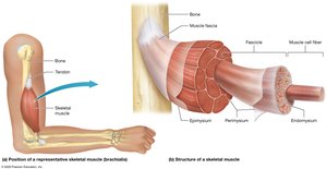
The skeletal muscle is a complex organ composed of muscle fibers, connective tissues, blood vessels, and nerves. Its organization allows for efficient force generation and movement.
Skeletal Muscle Fibers: Also known as skeletal muscle cells, these are long, thin cells surrounded by a thin layer of extracellular matrix called the endomysium.
Fascicle: A bundle of 10-100 muscle fibers, surrounded by connective tissue called the perimysium.
Epimysium: The outermost connective tissue sheath that surrounds the entire muscle, continuous with the fascia.
Tendons: Connect muscle to bone or other structures, transmitting the force generated by muscle contraction.
Blood and Nerve Supply: Skeletal muscles are richly supplied with blood vessels and nerves, essential for function and control.
Voluntary Control: Skeletal muscle contractions are under conscious control.
Fascicle Patterns and Muscle Shapes
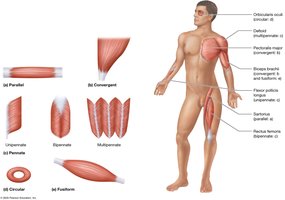
Muscles are classified by the arrangement of their fascicles and overall shape, which influences their function and range of motion.
Parallel: Fascicles are evenly spaced and run parallel to the muscle's long axis (e.g., sartorius).
Convergent: Broad at one end and tapers to a single tendon (e.g., pectoralis major).
Circular (Sphincters): Fascicles encircle an opening, closing it when contracted (e.g., orbicularis oculi).
Fusiform: Thick in the middle and tapered at both ends (e.g., biceps brachii).
Pennate: Fascicles attach to the tendon at an angle, resembling a feather. Types include:
Unipennate: Fascicles on one side of the tendon.
Bipennate: Fascicles on both sides of a single tendon.
Multipennate: Several tendons with fascicles arranged at multiple angles.
Naming Muscles
Muscles are named based on several criteria, which often describe their characteristics or location.
Size: (e.g., maximus, minimus, longus, brevis)
Location: (e.g., brachii for arm, femoris for thigh)
Shape: (e.g., deltoid for triangular shape)
Appearance: (e.g., striated, smooth)
Position: (e.g., lateralis for lateral, medialis for medial)
Number of Heads: (e.g., biceps for two heads, triceps for three)
Term | Meaning | Example |
|---|---|---|
Maximus | Largest | Gluteus maximus |
Minimus | Smallest | Gluteus minimus |
Longus | Long | Adductor longus |
Brevis | Short | Adductor brevis |
Biceps | Two heads | Biceps brachii |
Triceps | Three heads | Triceps brachii |
Femoris | Thigh | Quadriceps femoris |
Brachii | Arm | Biceps brachii |
Deltoid | Triangular | Deltoid |
Functions of Skeletal Muscles
Skeletal muscles perform essential functions for movement and homeostasis.
Muscle Tension: Generates force for body movements (actions) and heat production.
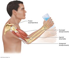
Functional Groups:
Agonists (Prime Movers): Main muscle responsible for a movement.
Antagonists: Oppose or slow the action of the agonist, allowing for controlled movement.
Synergists: Assist the agonist and help guide the movement.
Fixators: Stabilize the origin of the agonist, increasing efficiency and reducing injury risk.
Muscle Origin and Insertion
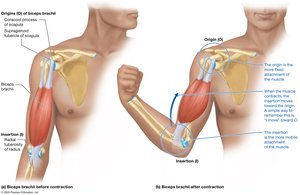
Muscles attach to bones at two main points, which determine the direction and type of movement produced.
Origin (O): The more fixed attachment point of the muscle.
Insertion (I): The attachment point that moves during contraction.
Example: The biceps brachii originates on the scapula (stationary) and inserts on the radius (moves).
For muscles crossing multiple joints, origin and insertion may switch depending on the movement.
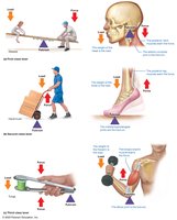
Lever Systems in Body Movements
Muscles and bones interact as lever systems to produce movement, with joints acting as fulcrums.
Lever: The rigid bar (bone) that moves.
Load: The object or body part being moved.
Force: The effort applied by muscle contraction.
Fulcrum: The pivot point (joint).
First-Class Lever: Fulcrum is between the load and force (e.g., nodding the head).
Second-Class Lever: Load is between the fulcrum and force (e.g., standing on tiptoe).
Third-Class Lever: Force is between the fulcrum and load (most common in the body, e.g., biceps curl).
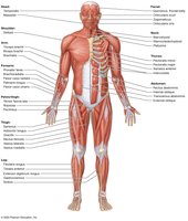
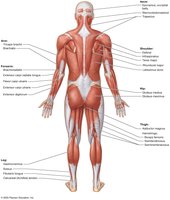
Superficial Muscles of the Body
The human body contains numerous superficial muscles, which are visible just beneath the skin and are responsible for major movements and body contours.
Anterior View: Includes muscles such as pectoralis major, rectus abdominis, quadriceps femoris, and biceps brachii.
Posterior View: Includes muscles such as trapezius, latissimus dorsi, gluteus maximus, and hamstrings.
Major Muscles and Their Actions
Muscles of Facial Expression
These muscles control facial movements and expressions.
Epicranius (Frontalis and Occipitalis): Elevates eyebrows and forehead skin.
Orbicularis Oculi: Closes eyelids (blinking, winking).
Zygomaticus Major and Minor: Assist with smiling.
Orbicularis Oris: Controls lips for eating, drinking, whistling, and speech.
Muscles of Mastication (Chewing)
Masseter: Elevates the mandible.
Temporalis: Elevates the mandible.
Muscles That Move the Head and Neck
Sternocleidomastoid: Rotates head to opposite shoulder; flexes head when both sides contract.
Trapezius (Superior part): Extends the head.
Platysma: Produces open-mouthed expression; tightens neck skin.
Muscles of Ventilation
Diaphragm: Main muscle for breathing; separates thoracic and abdominopelvic cavities.
External Intercostals: Raise and spread ribs for inspiration.
Internal Intercostals: Depress ribs for forced expiration.
Sternocleidomastoid: Assists in forced inspiration.
Abdominal Muscles
Rectus Abdominis: Flexes the trunk.
External/Internal Obliques: Rotate and laterally flex the trunk.
Transversus Abdominis: Compresses abdominal cavity, increases intra-abdominal pressure.
Functions: Facilitate urination, defecation, childbirth, and forced expiration.
Muscles That Move the Scapula (Pectoral Girdle)
Serratus Anterior: Protracts and rotates scapula superiorly.
Pectoralis Minor: Protracts and depresses scapula.
Trapezius: Elevates, retracts, and depresses scapula; assists with rotation.
Muscles That Move the Arm at the Shoulder Joint
Pectoralis Major: Flexes, adducts, and internally rotates arm.
Deltoid: Anterior fibers flex, central fibers abduct, and posterior fibers extend the arm.
Latissimus Dorsi: Extends, adducts, and internally rotates arm.
Teres Major: Assists latissimus dorsi.
Rotator Cuff Muscles: Stabilize shoulder joint; includes teres minor, supraspinatus, infraspinatus, subscapularis.
Muscles That Move the Forearm and Hand
Biceps Brachii: Flexes and supinates forearm.
Brachialis: Main elbow flexor.
Brachioradialis: Assists elbow flexion.
Triceps Brachii: Extends forearm; long head assists with adduction and stabilization.
Flexors (anterior/medial forearm): Flexor carpi radialis, palmaris longus, flexor carpi ulnaris, flexor digitorum superficialis.
Extensors (posterior/lateral forearm): Extensor carpi radialis longus, extensor carpi ulnaris, extensor digitorum.
Pronator Teres: Pronates forearm.
Muscles of the Hip, Thigh, Knee, and Leg
Iliopsoas: Main thigh flexor (iliacus + psoas major).
Pectineus: Assists thigh flexion and adduction.
Adductor Group: Adductor magnus, longus, brevis; adduct thigh.
Gracilis: Adducts thigh.
Sartorius: Flexes, abducts, laterally rotates thigh; flexes leg.
Quadriceps Femoris: Rectus femoris (flexes thigh, extends leg), vastus lateralis/intermedius/medialis (extend leg).
Gluteal Group: Gluteus maximus (extends, abducts, laterally rotates thigh), medius/minimus (abduct, medially rotate thigh).
Hamstrings: Semitendinosus, semimembranosus, biceps femoris (extend thigh and leg).
Muscles of the Ankle, Foot, and Toes
Tibialis Anterior, Extensor Digitorum Longus: Dorsiflex foot; tibialis anterior also inverts foot.
Fibularis Longus: Plantar flexion and eversion.
Gastrocnemius, Soleus: Plantar flex foot; gastrocnemius also flexes leg. Both form the calcaneal (Achilles) tendon.
