 Back
BackThe Muscular System: Structure, Function, and Major Muscles
Study Guide - Smart Notes
 Tailored notes based on your materials, expanded with key definitions, examples, and context.
The Muscular System
Introduction to the Muscular System
The muscular system is responsible for movement, posture, and heat production in the human body. It consists of skeletal muscles, which are under voluntary control, and is organized into functional groups based on their anatomical location and action.
Naming Muscles
Muscle Naming Conventions
Muscles are named according to several criteria, which help describe their characteristics and functions:
Size: Terms such as major, minor, longus (long), brevis (short), and vastus (broad) are used.
Location: Directional terms (e.g., superior, inferior, medial, lateral) and regional anatomical terms (e.g., pectoralis).
Attachment: Named for the bones or structures to which they attach (e.g., sternocleidomastoid).
Function: Named for their action, such as flexor, extensor, adductor, or abductor.
Number of Heads: Indicates the number of proximal attachments (e.g., biceps has two heads).
Functional Groups of Skeletal Muscles
Muscle Roles in Movement
Muscles work together to produce coordinated movements. They are classified into functional groups:
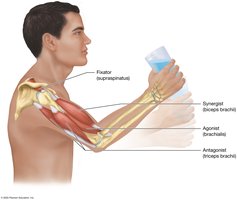
Agonist (Prime Mover): The main muscle responsible for a specific movement.
Antagonist: Lies on the opposite side of the joint from the agonist and opposes its action.
Synergist: Assists the agonist in performing its action.
Fixator: Stabilizes the origin of the agonist to allow efficient movement.
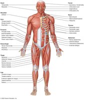
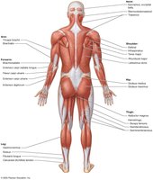
Overview of Major Muscle Groups
Muscle Group Classification
Muscles are categorized based on their anatomical location:
Muscles of the head, neck, and vertebral column
Muscles of the trunk and pelvic floor
Muscles of the pectoral girdle and upper limb
Muscles of the hip and lower limb
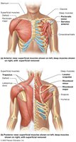
Familiarity with the superficial muscles (both anterior and posterior) is essential for understanding muscle function and identification.
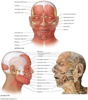
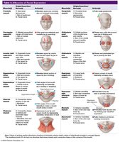
Muscles of Facial Expression
Key Muscles and Actions
Muscles of facial expression are responsible for movements of the face, such as smiling, frowning, and blinking:
Frontalis: Elevates the eyebrows.
Occipitalis: Pulls the scalp posteriorly.
Orbicularis oculi: Closes the eyelids (blinking/winking).
Zygomaticus major and minor: Elevate the corners of the mouth (smiling).
Orbicularis oris: Controls the lips for eating, drinking, and puckering.
Buccinator: Pulls the cheek inward (sucking).
Muscle | Origin/Insertion/Nerve | Action(s) |
|---|---|---|
Frontalis | Epicranial aponeurosis/skin of eyebrows/facial nerve | Raises eyebrows, wrinkles forehead |
Orbicularis oculi | Medial orbital margin/skin around eyelids/facial nerve | Closes eyelids |
Zygomaticus major | Zygomatic bone/skin at angle of mouth/facial nerve | Elevates corner of mouth |
Buccinator | Alveolar processes of maxilla and mandible/orbicularis oris/facial nerve | Compresses cheek |
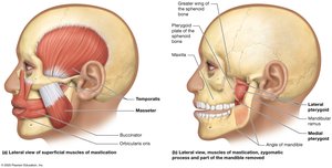
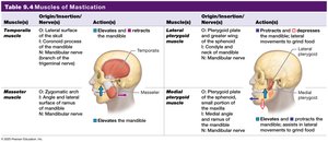
Muscles of Mastication
Key Muscles and Actions
Muscles of mastication are responsible for movements of the jaw during chewing:
Masseter: Elevates the mandible (closes the jaw).
Temporalis: Elevates the mandible.
Medial and Lateral Pterygoids: Move the mandible side-to-side and assist in grinding movements.
Muscle | Origin/Insertion/Nerve | Action(s) |
|---|---|---|
Temporalis | Temporal fossa/coronoid process of mandible/mandibular nerve | Elevates and retracts mandible |
Masseter | Zygomatic arch/mandibular ramus/mandibular nerve | Elevates mandible |
Lateral pterygoid | Lateral pterygoid plate/mandibular condyle/mandibular nerve | Protracts and depresses mandible |
Medial pterygoid | Medial pterygoid plate/mandibular angle/mandibular nerve | Elevates and protracts mandible |
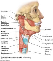
Muscles of Swallowing
Key Muscles and Actions
Muscles involved in swallowing coordinate the movement of the hyoid bone and larynx:
Omohyoid and Sternohyoid: Depress the hyoid bone during swallowing.
Mylohyoid, Geniohyoid, Stylohyoid: Elevate the hyoid bone and floor of the mouth.
Muscle | Origin/Insertion/Nerve | Action(s) |
|---|---|---|
Omohyoid | Superior border of scapula/hyoid bone/ansa cervicalis | Depresses hyoid bone |
Sternohyoid | Manubrium/hyoid bone/ansa cervicalis | Depresses hyoid bone |
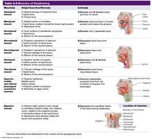
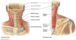
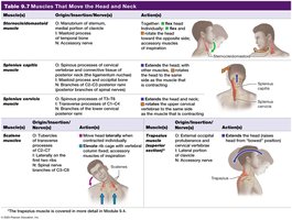
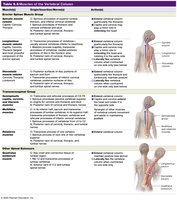
Muscles That Move the Head and Neck
Key Muscles and Actions
These muscles are responsible for movements such as rotation, flexion, and extension of the head and neck:
Sternocleidomastoid: Rotates the head laterally and flexes the neck.
Trapezius: Extends the head and neck.
Splenius capitis: Rotates and extends the head and neck.
Muscle | Origin/Insertion/Nerve | Action(s) |
|---|---|---|
Sternocleidomastoid | Sternum and clavicle/mastoid process/accessory nerve | Flexes and rotates head |
Trapezius | Occipital bone, spinous processes of C7-T12/clavicle, scapula/accessory nerve | Extends head and neck |
Splenius capitis | Spinous processes of cervical vertebrae/mastoid process/cervical spinal nerves | Extends, rotates head |
Muscles of the Vertebral Column
Key Muscles and Actions
These muscles maintain posture and allow movement of the vertebral column:
Erector spinae group: Extends and laterally flexes the vertebral column.
Longissimus thoracis: Assists in extending or rotating the head and neck.
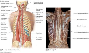
Muscle | Origin/Insertion/Nerve | Action(s) |
|---|---|---|
Erector spinae | Iliac crest, sacrum, lumbar vertebrae/ribs, thoracic and cervical vertebrae/spinal nerves | Extends vertebral column |
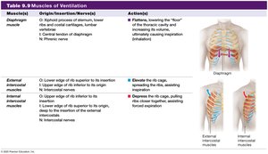
Muscles of Ventilation
Key Muscles and Actions
Muscles of ventilation are essential for breathing by changing the volume of the thoracic cavity:
Diaphragm: Contracts to increase thoracic volume during inspiration.
External intercostals: Elevate the ribs during inspiration.
Internal intercostals: Depress the ribs during forced exhalation.
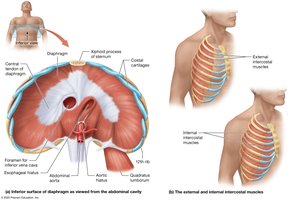
Muscle | Origin/Insertion/Nerve | Action(s) |
|---|---|---|
Diaphragm | Xiphoid process, lower ribs/central tendon/phrenic nerve | Increases thoracic cavity volume (inhalation) |
External intercostals | Inferior border of rib above/superior border of rib below/intercostal nerves | Elevate ribs (inspiration) |
Internal intercostals | Superior border of rib below/inferior border of rib above/intercostal nerves | Depress ribs (forced expiration) |
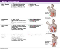
Abdominal Muscles
Key Muscles and Actions
Abdominal muscles support the trunk, allow movement, and hold organs in place:
Rectus abdominis: Compresses the abdomen and flexes the trunk.
External and internal obliques: Rotate and laterally flex the trunk.
Transversus abdominis: Compresses the abdominal cavity.
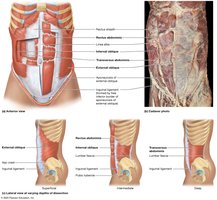
Muscle | Origin/Insertion/Nerve | Action(s) |
|---|---|---|
Rectus abdominis | Pubic crest, symphysis/xiphoid process, costal cartilages/thoracoabdominal nerves | Flexes trunk, compresses abdomen |
External oblique | Lower eight ribs/iliac crest, linea alba/thoracoabdominal nerves | Flexes, rotates trunk |
Transversus abdominis | Lower six ribs, iliac crest/linea alba/thoracoabdominal nerves | Compresses abdomen |
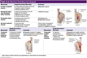
Muscles That Move the Scapula at the Pectoral Girdle
Key Muscles and Actions
These muscles stabilize and move the scapula, allowing for a wide range of shoulder movements:
Serratus anterior: Protracts the scapula.
Pectoralis minor: Depresses the scapula.
Trapezius: Extends the head and neck, elevates and retracts the scapula.
Rhomboid major and minor: Retract the scapula.
Muscle | Origin/Insertion/Nerve | Action(s) |
|---|---|---|
Serratus anterior | Ribs 1-8/medial border of scapula/long thoracic nerve | Protracts scapula |
Pectoralis minor | Ribs 3-5/coracoid process/medial pectoral nerve | Depresses scapula |
Trapezius | Occipital bone, spinous processes of C7-T12/clavicle, scapula/accessory nerve | Elevates, retracts scapula |
Rhomboid major/minor | Spinous processes of C7-T5/medial border of scapula/dorsal scapular nerve | Retracts scapula |
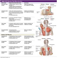
Muscles That Move the Arm at the Shoulder Joint
Key Muscles and Actions
These muscles are responsible for movements of the arm at the shoulder joint:
Pectoralis major: Adducts the arm.
Coracobrachialis: Assists with flexion of the humerus.
Deltoid: Abducts the arm.
Latissimus dorsi: Extends the arm.
Teres major: Extends and adducts the arm.
Rotator cuff muscles: Stabilize and move the shoulder joint (includes supraspinatus, infraspinatus, teres minor, subscapularis).
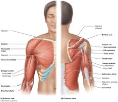
Muscle | Origin/Insertion/Nerve | Action(s) |
|---|---|---|
Pectoralis major | Clavicle, sternum/humerus/medial and lateral pectoral nerves | Adducts, flexes arm |
Deltoid | Clavicle, scapula/humerus/axillary nerve | Abducts arm |
Latissimus dorsi | Spinous processes of lower thoracic vertebrae/humerus/thoracodorsal nerve | Extends, adducts arm |
Rotator cuff muscles | Scapula/humerus/suprascapular, axillary, subscapular nerves | Stabilize shoulder |
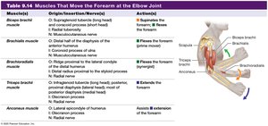
Muscles That Move the Forearm and Hand
Muscles That Move the Forearm at the Elbow Joint
Biceps brachii, brachialis, brachioradialis: Flex the forearm.
Triceps brachii: Extends the forearm.
Muscle | Origin/Insertion/Nerve | Action(s) |
|---|---|---|
Biceps brachii | Scapula/radius/musculocutaneous nerve | Flexes forearm, supinates forearm |
Brachialis | Humerus/ulna/musculocutaneous nerve | Flexes forearm |
Brachioradialis | Humerus/radius/radial nerve | Flexes forearm |
Triceps brachii | Scapula, humerus/ulna/radial nerve | Extends forearm |
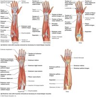
Muscles That Move the Hand and Fingers
Flexors: Located on the anterior and medial forearm; flex the hand (e.g., flexor carpi radialis, palmaris longus, flexor carpi ulnaris).
Pronator teres: Pronates the forearm.
Extensors: Located on the posterior and lateral forearm; extend the hand and wrist (e.g., extensor carpi radialis longus, extensor carpi ulnaris, extensor digitorum).
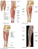
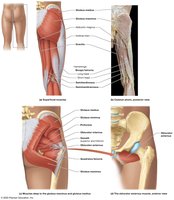
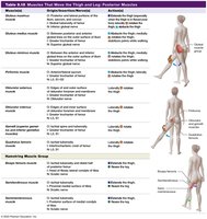
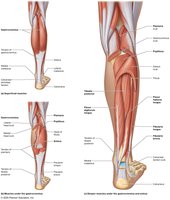
Muscles of the Hip, Thigh, Knee, and Leg
Anterior and Medial Muscles
Iliopsoas (iliacus and psoas major): Flex the thigh.
Adductor group (magnus, longus, brevis): Adduct the thigh.
Gracilis: Adducts the thigh.
Sartorius: Flexion and lateral rotation of the thigh.
Quadriceps femoris group (rectus femoris, vastus lateralis, intermedius, medialis): Extend the leg at the knee joint.
Muscle | Origin/Insertion/Nerve | Action(s) |
|---|---|---|
Iliopsoas | Iliac fossa, lumbar vertebrae/femur/femoral nerve | Flexes thigh |
Adductor magnus | Ischial tuberosity/femur/obturator nerve | Adducts thigh |
Quadriceps femoris | Ilium, femur/patella, tibia/femoral nerve | Extends leg |
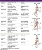

Posterior Muscles
Gluteal group (maximus, medius, minimus): Extend and abduct the thigh.
Piriformis: Laterally rotates the thigh.
Hamstring group (semitendinosus, semimembranosus, biceps femoris): Extend the thigh and flex the leg.
Muscle | Origin/Insertion/Nerve | Action(s) |
|---|---|---|
Gluteus maximus | Ilium, sacrum/femur/inferior gluteal nerve | Extends thigh |
Hamstrings | Ischial tuberosity/tibia, fibula/sciatic nerve | Extends thigh, flexes leg |
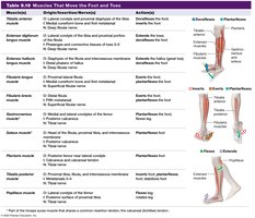
Muscles of the Ankle, Foot, and Toes
Key Muscles and Actions
Tibialis anterior, extensor digitorum longus: Dorsiflexion of the foot.
Fibularis longus: Plantarflexion and eversion of the foot.
Gastrocnemius, soleus: Plantarflexion of the foot.
Flexor digitorum longus: Flexes the digits (toes).
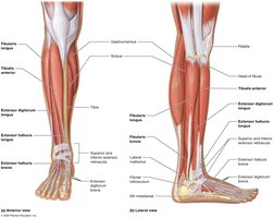
Muscle | Origin/Insertion/Nerve | Action(s) |
|---|---|---|
Tibialis anterior | Tibia/medial cuneiform, first metatarsal/deep fibular nerve | Dorsiflexes foot |
Gastrocnemius | Femur/calcaneus/tibial nerve | Plantarflexes foot |
Flexor digitorum longus | Tibia/distal phalanges of toes/tibial nerve | Flexes toes |
Additional info: For each muscle group, understanding the origin, insertion, innervation, and action is essential for clinical and academic applications. Tables referenced above provide detailed anatomical and functional information for each muscle.
