Back
BackThe Muscular System: Structure, Function, and Physiology
Study Guide - Smart Notes
 Tailored notes based on your materials, expanded with key definitions, examples, and context.
The Muscular System
Overview of Muscle Types
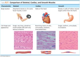
The muscular system is responsible for all types of body movement and is composed of three basic muscle types: skeletal muscle, cardiac muscle, and smooth muscle. Each type has distinct structural and functional characteristics that enable them to perform specialized roles in the body.
Skeletal muscle: Primarily attached to bones and responsible for voluntary movements.
Cardiac muscle: Found only in the heart, responsible for pumping blood.
Smooth muscle: Located in the walls of hollow organs, responsible for involuntary movements such as peristalsis.
Muscle Types
Skeletal Muscle
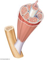
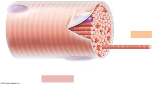
Skeletal muscle fibers are large, cigar-shaped, and multinucleate. They are also known as striated muscle due to their striped appearance and as voluntary muscle because they are under conscious control. Most skeletal muscles are attached to bones by tendons, which are tough, cordlike structures composed mainly of collagen fibers. In some cases, muscles are attached by aponeuroses, which are sheetlike structures that connect muscles to bones, cartilage, or connective tissue coverings.
Cell shape: Long, cylindrical, multinucleate, with obvious striations.
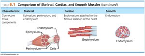
Connective tissue wrappings: Each muscle fiber is wrapped in endomysium, fascicles are wrapped in perimysium, and the entire muscle is wrapped in epimysium.
Smooth Muscle
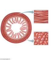
Smooth muscle lacks striations and is involuntary, meaning it is not under conscious control. It is found mainly in the walls of hollow visceral organs such as the stomach, urinary bladder, and respiratory passages. Smooth muscle fibers are spindle-shaped, uninucleate, and contract slowly and in a sustained manner.
Cell shape: Single, fusiform, uninucleate, no striations.
Location: Walls of hollow organs (other than the heart).
Cardiac Muscle
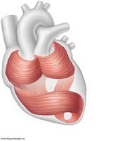
Cardiac muscle is found only in the heart. It is striated and involuntary. Cardiac muscle cells are branching, uninucleate, and joined by gap junctions called intercalated discs. The heart contracts at a steady rate set by a pacemaker.
Cell shape: Branching chains of cells, uninucleate, striations, intercalated discs.
Location: Walls of the heart.
Microscopic Anatomy of Skeletal Muscle
Muscle Fiber Structure
Skeletal muscle fibers contain specialized structures that enable contraction. The sarcolemma is the specialized plasma membrane, and myofibrils are long organelles inside the muscle cell. The alternating light (I) and dark (A) bands give the muscle its striated appearance.
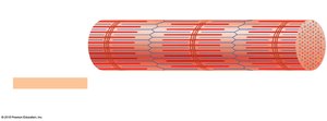
Sarcomere: The Contractile Unit
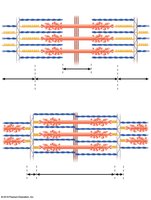
The sarcomere is the structural and functional unit of skeletal muscle. It is composed of thick (myosin) and thin (actin) filaments, which produce the banding pattern seen in striated muscle. The arrangement of these filaments allows for muscle contraction through the sliding filament mechanism.
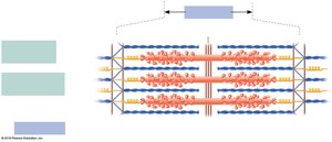
Sarcoplasmic Reticulum (SR)
The sarcoplasmic reticulum is a specialized smooth endoplasmic reticulum that surrounds the myofibril and stores and releases calcium ions, which are essential for muscle contraction.
Stimulation and Contraction of Skeletal Muscle
Functional Properties
Irritability (Responsiveness): Ability to receive and respond to a stimulus.
Contractility: Ability to forcibly shorten when an adequate stimulus is received.
Extensibility: Ability of muscle cells to be stretched.
Elasticity: Ability to recoil and resume resting length after stretching.
The Nerve Stimulus and Action Potential
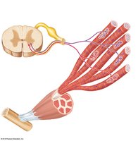
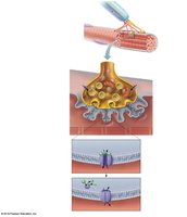
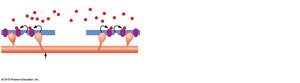
Skeletal muscles must be stimulated by a motor neuron to contract. A motor unit consists of one motor neuron and all the skeletal muscle cells it stimulates. The neuromuscular junction is the association site of the axon terminal of the motor neuron and the sarcolemma of a muscle fiber. The neurotransmitter acetylcholine (ACh) is released upon nerve impulse arrival, initiating muscle contraction.
Mechanism of Muscle Contraction
Sliding Filament Theory
Muscle contraction occurs when calcium ions bind to regulatory proteins on actin filaments, exposing myosin-binding sites. Myosin heads attach to these sites, forming cross-bridges and pulling the actin filaments toward the center of the sarcomere, resulting in contraction.
Energy for Muscle Contraction
ATP and Muscle Contraction
ATP is the only energy source that can be used directly to power muscle contraction. Because stored ATP is limited, muscle fibers regenerate ATP through three main pathways:
Direct phosphorylation: Creatine phosphate (CP) donates a phosphate to ADP to form ATP. This process is anaerobic and provides energy for about 15 seconds.
Aerobic respiration: Occurs in mitochondria, uses oxygen, and produces 32 ATP per glucose molecule. It is the primary energy source for prolonged, moderate activity.
Anaerobic glycolysis: Occurs in the cytosol, does not require oxygen, and produces 2 ATP per glucose molecule along with lactic acid. It provides energy for short bursts of activity.
Muscle Fatigue and Oxygen Deficit
Muscle fatigue occurs after prolonged or strenuous activity due to factors such as ion imbalances, oxygen deficit, lactic acid accumulation, and decreased ATP supply. After exercise, the oxygen deficit is repaid by rapid, deep breathing.
Types of Muscle Contractions
Isotonic contractions: Myofilaments slide past each other, the muscle shortens, and movement occurs (e.g., bending the knee, lifting weights).
Isometric contractions: Muscle filaments attempt to slide, but the muscle is pitted against an immovable object, so tension increases but the muscle does not shorten (e.g., pushing palms together).
Muscle Tone
Muscle tone is the state of continuous partial contractions, resulting from systematic stimulation of different motor units. It keeps muscles firm, healthy, and ready for action.
Effect of Exercise on Muscles
Exercise increases muscle size, strength, and endurance. Aerobic (endurance) exercise improves muscle flexibility, resistance to fatigue, metabolism, digestion, and coordination. Resistance (isometric) exercise increases muscle size and strength by enlarging individual muscle fibers.