 Back
The Muscular System: Structure, Function, and Types of Muscle Tissue
Study Guide - Smart Notes
 Tailored notes based on your materials, expanded with key definitions, examples, and context.
The Muscular System
Overview of Muscle Tissues
The human body contains three main types of muscle tissue: skeletal, cardiac, and smooth muscle. Each type has distinct structural and functional characteristics that enable the body to perform a wide range of movements and physiological processes.
Skeletal muscle: Voluntary muscles attached to bones, responsible for body movement.
Cardiac muscle: Involuntary muscle found only in the heart, responsible for pumping blood.
Smooth muscle: Involuntary muscle found in the walls of hollow organs, responsible for movements such as peristalsis in the digestive tract.
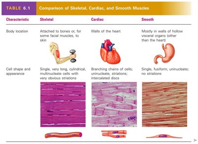
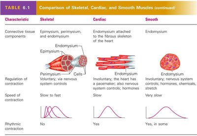
Comparison of Muscle Types
The following table summarizes the main differences between skeletal, cardiac, and smooth muscle tissues:
Characteristic | Skeletal | Cardiac | Smooth |
|---|---|---|---|
Location | Attached to bones or skin | Walls of the heart | Walls of hollow organs (except heart) |
Cell Shape & Appearance | Long, cylindrical, multinucleate, striated | Branching chains, uninucleate, striated, intercalated discs | Fusiform, uninucleate, non-striated |
Control | Voluntary | Involuntary | Involuntary |
Speed of Contraction | Slow to fast | Slow | Very slow |
Rhythmic Contraction | No | Yes | Yes, in some |
Connective Tissue Components of Skeletal Muscle
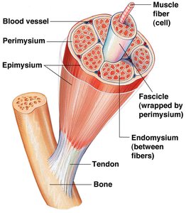
Skeletal muscle fibers are organized and supported by several layers of connective tissue:
Endomysium: Surrounds each individual muscle fiber (cell).
Perimysium: Surrounds bundles of muscle fibers called fascicles.
Epimysium: Encloses the entire muscle.
Tendon: Connects muscle to bone, formed by the merging of connective tissue layers.
Microscopic Anatomy of Muscle Tissue
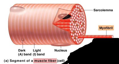
Muscle fibers contain specialized structures for contraction:
Sarcolemma: The plasma membrane of a muscle cell.
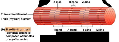
Myofibrils: Long, rod-like organelles within muscle fibers, composed of repeating units called sarcomeres.
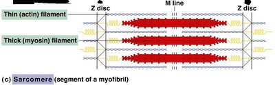
Sarcomere: The functional contractile unit of muscle, defined by Z discs at each end.
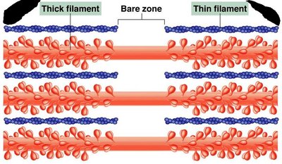
Myofilaments: Thin (actin) and thick (myosin) filaments within the sarcomere responsible for muscle contraction.
Arrangement of Smooth and Cardiac Muscle
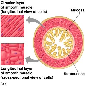
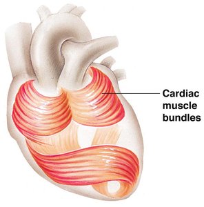
Smooth muscle is typically arranged in two layers (circular and longitudinal) in the walls of hollow organs, allowing for coordinated contractions such as peristalsis. Cardiac muscle fibers are arranged in spiral or figure-8 bundles, optimizing the heart's pumping action.
Banding Pattern and Zones in Skeletal Muscle
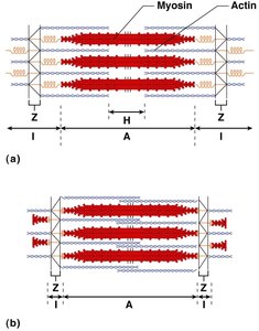
The striated appearance of skeletal muscle is due to the arrangement of myofilaments:
I band: Light band, contains only thin filaments (actin).
A band: Dark band, contains the entire length of thick filaments (myosin).
H zone: Central region of the A band, contains only thick filaments.
Z disc: Defines the boundary of each sarcomere, anchors thin filaments.
M line: Center of the H zone, holds thick filaments together.
The Sliding Filament Theory of Muscle Contraction
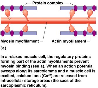
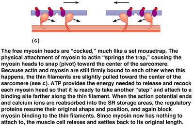
Muscle contraction occurs when myosin heads bind to actin filaments and pull them toward the center of the sarcomere, shortening the muscle fiber. This process is powered by ATP and regulated by calcium ions released from the sarcoplasmic reticulum.
In a relaxed muscle, regulatory proteins block myosin binding sites on actin.
Upon stimulation, calcium ions bind to regulatory proteins, exposing binding sites for myosin.
Myosin heads attach, pivot, and pull actin filaments inward (power stroke).
ATP is required to detach and reset the myosin heads for another cycle.
Summary Table: Key Differences Between Muscle Types
Feature | Skeletal Muscle | Cardiac Muscle | Smooth Muscle |
|---|---|---|---|
Location | Bones, skin | Heart | Walls of hollow organs |
Control | Voluntary | Involuntary | Involuntary |
Striations | Yes | Yes | No |
Cell Shape | Long, cylindrical, multinucleate | Branched, uninucleate | Fusiform, uninucleate |
Speed of Contraction | Slow to fast | Slow | Very slow |
Rhythmic Contraction | No | Yes | Yes, in some |
Additional info: The arrangement of muscle fibers and connective tissue layers is essential for the transmission of force and the overall function of muscle tissue. The sliding filament theory is fundamental to understanding how muscles contract and generate movement.
