 Back
The Muscular System: Structure, Function, and Major Muscles
Study Guide - Smart Notes
 Tailored notes based on your materials, expanded with key definitions, examples, and context.
The Muscular System
Introduction to the Muscular System
The muscular system is essential for movement, posture, and various physiological processes. It consists of three main types of muscle tissue: skeletal, cardiac, and smooth muscle. This chapter focuses on skeletal muscles, which are responsible for voluntary movements and are attached to bones via tendons.
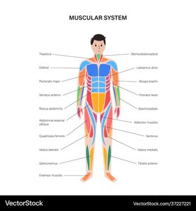
Muscle Actions and Interactions
Prime Movers (Agonists), Antagonists, and Synergists
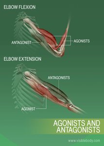
Muscles work together to produce coordinated movements. Each muscle can play different roles depending on the movement:
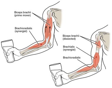
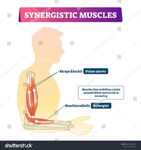
Prime Mover (Agonist): The main muscle responsible for a specific movement. Example: Biceps brachii during elbow flexion.
Antagonist: Muscle that opposes or reverses the action of the prime mover. Example: Triceps brachii during elbow flexion.
Synergist: Muscle that assists the prime mover by adding force or reducing unwanted movements. Example: Brachialis assists the biceps brachii.
Fixator: A type of synergist that stabilizes the origin of the prime mover.
Skeletal Muscle Attachments: Origin and Insertion
Definitions and Functional Importance
Muscles attach to bones at two main points:
Origin: The attachment site on the bone that moves the least during contraction (usually proximal).
Insertion: The attachment site on the bone that moves the most during contraction (usually distal).
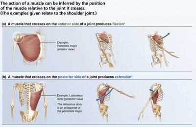
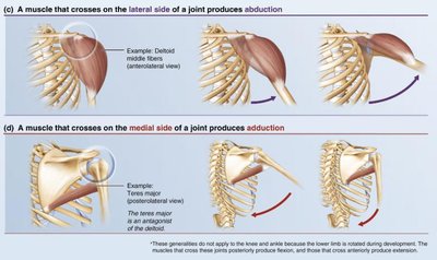
The action of a muscle is determined by its position relative to the joint it crosses:
Anterior to joint: typically causes flexion
Posterior to joint: typically causes extension
Lateral to joint: typically causes abduction
Medial to joint: typically causes adduction
Criteria for Naming Muscles
Muscle Naming Conventions
Muscles are named based on several criteria, which often describe their characteristics or location:
Location: e.g., Tibialis anterior (near the tibia, anterior side of leg)
Size: e.g., Gluteus maximus (largest in gluteal region)
Shape: e.g., Deltoid (triangular shape)
Action: e.g., Flexor carpi ulnaris (flexes wrist, near ulna)
Number of origins: e.g., Biceps (two origins), Triceps (three origins)
Direction of fibers: e.g., Rectus (straight), Transversus (right angles), Oblique (diagonal)
Muscle Fascicle Arrangement
Patterns and Functional Implications
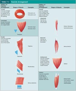
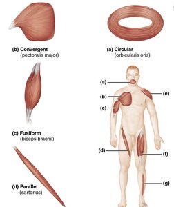
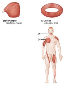
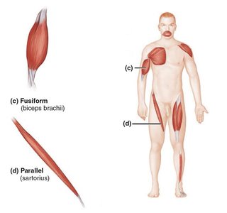
Muscle fascicles are bundles of muscle fibers arranged in specific patterns, influencing power and range of motion:
Parallel: Fascicles run parallel to the long axis (e.g., sartorius). Greater range of motion, less power.
Fusiform: Spindle-shaped with parallel fibers (e.g., biceps brachii).
Circular: Fascicles arranged in concentric rings (e.g., orbicularis oris). Act as sphincters.
Convergent: Broad origin, fascicles converge toward a single tendon (e.g., pectoralis major).
Pennate: Short fascicles attach obliquely to a central tendon. Types: unipennate, bipennate, multipennate (e.g., rectus femoris, deltoid).
Levers and Biomechanics in Muscle Action
Definition and Principles of Levers
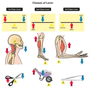
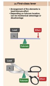
A lever is a rigid bar that moves on a fixed point called a fulcrum. In the body, bones act as levers, joints as fulcrums, and muscles provide the effort to move a load. Levers can operate at a mechanical advantage (power lever) or disadvantage (speed lever):
Mechanical Advantage: Load is closer to fulcrum than effort; less effort needed, but slower movement.
Mechanical Disadvantage: Effort is closer to fulcrum than load; more effort needed, but faster movement.
Types of Lever Systems
Class | Arrangement | Example | Advantage |
|---|---|---|---|
First Class | Fulcrum between effort and load (E-F-L) | Atlanto-occipital joint (nodding head) | Can be advantage or disadvantage |
Second Class | Load between fulcrum and effort (F-L-E) | Standing on tiptoes | Always advantage (powerful) |
Third Class | Effort between fulcrum and load (F-E-L) | Biceps curl | Always disadvantage (speed/range) |
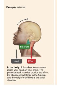
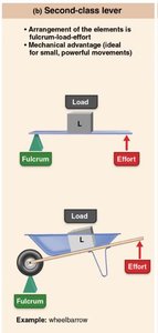
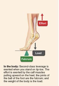
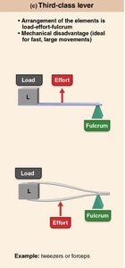
Major Skeletal Muscles of the Body
Overview and Identification
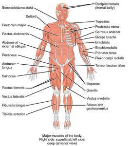
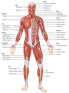
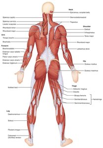
There are over 600 skeletal muscles, grouped by function and location. Understanding their origin, insertion, and action is fundamental for analyzing movement and diagnosing musculoskeletal issues.
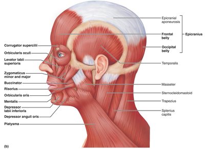
Muscles of Facial Expression
Unique Features and Examples
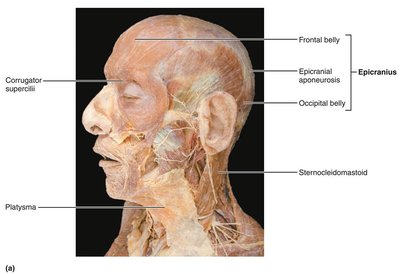
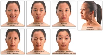
Facial muscles are unique because they insert into the skin rather than bone, allowing for a wide range of expressions. All are innervated by the facial nerve (cranial nerve VII).
Muscle | Description | Origin/Insertion | Action | Nerve Supply |
|---|---|---|---|---|
Epicranius (frontal belly) | Covers forehead | O: epicranial aponeurosis I: skin of eyebrows | Raises eyebrows, wrinkles forehead | Facial nerve (VII) |
Orbicularis oculi | Sphincter of eyelid | O: frontal/maxillary bones I: tissue of eyelid | Closes eye, blinking | Facial nerve (VII) |
Zygomaticus | Extends from cheekbone to mouth | O: zygomatic bone I: skin at mouth corner | Raises mouth corners (smile) | Facial nerve (VII) |
Orbicularis oris | Encircles mouth | O: maxilla/mandible I: skin at mouth angles | Closes, protrudes lips | Facial nerve (VII) |
Buccinator | Cheek muscle | O: molar region of maxilla/mandible I: orbicularis oris | Compresses cheek | Facial nerve (VII) |
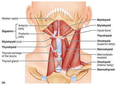
Muscles of the Anterior Neck and Throat: Swallowing
Suprahyoid and Infrahyoid Muscles
These muscles are involved in swallowing and speech. The suprahyoid group elevates the hyoid bone and larynx, while the infrahyoid group depresses them after swallowing. The pharyngeal constrictor muscles propel food into the esophagus.
Disorders and Health of the Muscular System
Common Disorders
Muscular Dystrophy: Inherited disorder causing muscle degeneration and weakness.
Duchenne Muscular Dystrophy: Caused by lack of a protein to maintain the sarcolemma.
Myasthenia Gravis: Progressive weakness due to a shortage of acetylcholine receptors.
Muscle Fatigue and Recovery
Muscle fatigue is the decline in force production during prolonged activity.
Recovery involves replenishing energy stores, repairing muscle damage, and removing metabolic waste.
Muscle Injuries and Prevention
Common injuries: strains, sprains, contusions.
Prevention: proper warm-up, stretching, and strength training.
Role of Nutrition in Muscle Health
Adequate protein for repair and growth.
Carbohydrates for energy.
Vitamins and minerals for muscle function.
