 Back
The Muscular System: Structure, Function, and Physiology
Study Guide - Smart Notes
 Tailored notes based on your materials, expanded with key definitions, examples, and context.
The Muscular System
Overview of Muscle Types
The human body contains three primary types of muscle tissue: skeletal muscle, cardiac muscle, and smooth muscle. Each type has distinct structural and functional characteristics that enable the body to perform a wide range of movements and physiological processes.
Skeletal muscle: Responsible for voluntary movements and attached to bones.
Cardiac muscle: Found only in the heart, responsible for pumping blood.
Smooth muscle: Located in the walls of hollow organs, responsible for involuntary movements such as peristalsis.
Comparison of Muscle Types
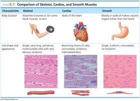
The following table summarizes the main differences among skeletal, cardiac, and smooth muscle tissues, including their location, structure, and function.
Characteristic | Skeletal | Cardiac | Smooth |
|---|---|---|---|
Body Location | Attached to bones or some facial muscles | Walls of the heart | Walls of hollow visceral organs (except heart) |
Cell Shape & Appearance | Single, very long, cylindrical, multinucleate, striated | Branching chains, uninucleate, striated, intercalated discs | Single, fusiform, uninucleate, no striations |
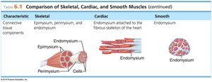
Connective Tissue Components | Epimysium, perimysium, endomysium | Endomysium attached to fibrous skeleton of heart | Endomysium |
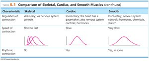
Regulation of Contraction | Voluntary (nervous system controls) | Involuntary (pacemaker, nervous system, hormones) | Involuntary (nervous system, hormones, chemicals, stretch) |
Speed of Contraction | Slow to fast | Slow | Very slow |
Rhythmic Contraction | No | Yes | Yes, in some |
Structure of Skeletal Muscle
Connective Tissue Wrappings
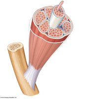
Skeletal muscle fibers are organized and protected by several layers of connective tissue:
Endomysium: Surrounds each individual muscle fiber.
Perimysium: Encloses a bundle of muscle fibers (fascicle).
Epimysium: Encloses the entire muscle.
Fascia: Outermost layer, separates muscles from surrounding tissues.
Attachment to Bones
The epimysium blends into connective tissue attachments:
Tendons: Cordlike structures that attach muscle to bone, composed mainly of collagen fibers.
Aponeuroses: Sheetlike structures that attach muscles indirectly to bones or other connective tissues.
Microscopic Anatomy of Skeletal Muscle
Muscle Fiber Structure
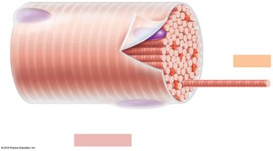
Each skeletal muscle fiber (cell) contains specialized structures for contraction:
Sarcolemma: The plasma membrane of a muscle fiber.
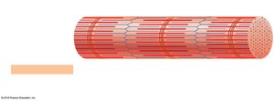
Myofibrils: Long, rod-like organelles that fill the muscle fiber and are responsible for the striated appearance.
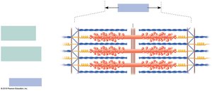
Sarcomere: The contractile unit of a muscle fiber, defined as the region between two Z discs.
Myofilaments
Myofibrils are composed of two main types of myofilaments:
Thick filaments (myosin): Contain myosin heads that form cross-bridges during contraction.
Thin filaments (actin): Anchored to the Z disc; interact with myosin for muscle contraction.
The arrangement of these filaments creates the characteristic banding pattern of skeletal muscle.
Sarcoplasmic Reticulum (SR)
The SR is a specialized smooth endoplasmic reticulum that stores and releases calcium ions, which are essential for muscle contraction.
Muscle Contraction: The Sliding Filament Theory
Mechanism of Contraction
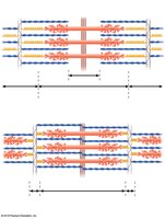
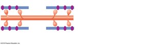
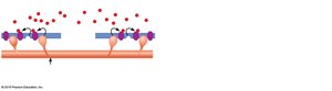
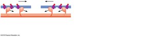
Muscle contraction occurs when myosin heads bind to actin, forming cross-bridges and pulling the thin filaments toward the center of the sarcomere. This process shortens the sarcomere and, consequently, the muscle fiber.
Calcium ions bind to regulatory proteins on actin, exposing myosin-binding sites.
ATP provides the energy for myosin head movement and detachment.
Stimulation and Contraction of Skeletal Muscle Cells
Neuromuscular Junction and Action Potential
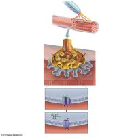
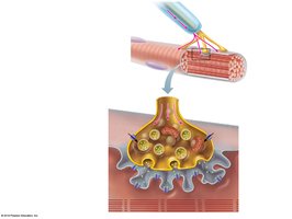
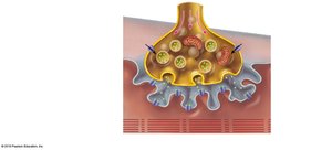
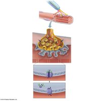
Skeletal muscle fibers contract in response to stimulation by motor neurons at the neuromuscular junction. The process involves the following steps:
Nerve impulse reaches the axon terminal of the motor neuron.
Calcium channels open, allowing Ca2+ to enter the terminal.
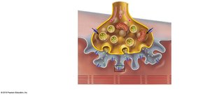
Synaptic vesicles release acetylcholine (ACh) into the synaptic cleft.
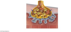
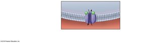
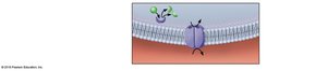
ACh binds to receptors on the sarcolemma, increasing permeability to Na+ and K+.
Depolarization occurs, generating an action potential that spreads along the muscle fiber.
Acetylcholinesterase breaks down ACh, ending the signal and allowing the muscle to relax.
Energy for Muscle Contraction
ATP and Muscle Contraction
ATP is the only direct energy source for muscle contraction. Because muscle fibers store limited ATP, they must regenerate it through three main pathways:
Direct phosphorylation by creatine phosphate (CP): Fastest, provides energy for about 15 seconds.
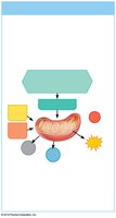
Aerobic respiration: Occurs in mitochondria, uses oxygen, produces about 32 ATP per glucose, supports prolonged activity.
Anaerobic glycolysis: Occurs in cytosol, does not require oxygen, produces 2 ATP per glucose and lactic acid, supports short bursts of activity.
Types of Muscle Contractions
Isotonic vs. Isometric Contractions
Isotonic contractions: Muscle changes length and movement occurs (e.g., lifting a weight).
Isometric contractions: Muscle develops tension but does not change length (e.g., pushing against an immovable object).
Effect of Exercise on Muscles
Aerobic vs. Resistance Training
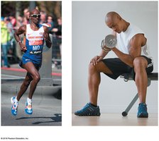
Aerobic (endurance) exercise: Increases muscle endurance, flexibility, and resistance to fatigue; improves metabolism and coordination.
Resistance (isometric) exercise: Increases muscle size and strength by enlarging individual muscle fibers.
Summary Table: Muscle Types
Feature | Skeletal Muscle | Cardiac Muscle | Smooth Muscle |
|---|---|---|---|
Location | Bones | Heart | Hollow organs |
Control | Voluntary | Involuntary | Involuntary |
Striations | Yes | Yes | No |
Cell Shape | Long, cylindrical, multinucleate | Branched, uninucleate | Fusiform, uninucleate |
Contraction Speed | Slow to fast | Slow | Very slow |
