 Back
The Muscular System: Structure, Function, and Disorders
Study Guide - Smart Notes
 Tailored notes based on your materials, expanded with key definitions, examples, and context.
Muscular System Overview
Introduction to Muscle Tissue
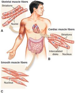
The muscular system enables movement of the body and its parts through the contraction of muscle cells, also known as fibers. Muscle cells convert chemical energy from food into mechanical energy, resulting in movement. There are three main types of muscle tissue in the human body: skeletal, cardiac, and smooth muscle.
Skeletal muscle: Attaches to bones, also called striated or voluntary muscle due to its striped appearance and voluntary control.
Cardiac muscle: Found only in the heart, characterized by branching cells and intercalated disks, allowing the heart to contract as a unit.
Smooth muscle: Located in the walls of hollow organs, lacks striations, and is under involuntary control.
Skeletal Muscle Structure and Function
Macroscopic and Microscopic Structure
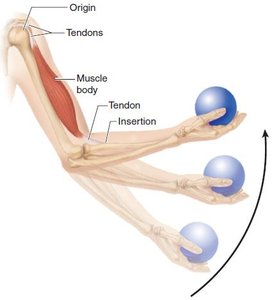
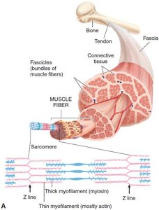
Skeletal muscles are organs composed of muscle fibers and connective tissue. Each muscle fiber is wrapped in fibrous connective tissue, which continues around groups of fibers (fascicles) and the entire muscle. Muscles attach to bones via tendons, and movement occurs across joints.
Origin: Attachment to the stationary bone.
Insertion: Attachment to the bone that moves during contraction.
Body: The main part of the muscle.
Microscopic Structure
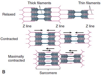
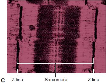
Muscle fibers contain bundles of myofilaments: thick filaments (myosin) and thin filaments (actin). The basic contractile unit is the sarcomere, separated by Z lines. The sliding filament model explains muscle contraction, where myofilaments slide past each other, shortening the sarcomere.
Mechanism of Muscle Contraction
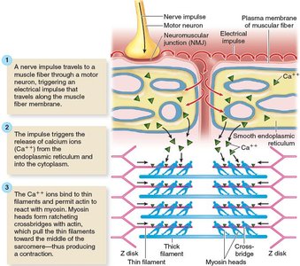
Muscle contraction is initiated by a nerve impulse at the neuromuscular junction (NMJ), leading to the release of calcium ions and ATP, which are essential for the sliding of actin and myosin filaments.
Calcium ions bind to thin filaments, allowing myosin heads to form cross-bridges with actin.
ATP is required for both contraction and relaxation of muscle fibers.
Muscle Function and Movement
Types of Muscle Contraction
Muscles produce movement by pulling on bones. The type of contraction determines the nature of the movement:
Tonic contraction: Maintains posture without producing movement.
Twitch contraction: Quick, jerky response to a stimulus.
Tetanic contraction: Sustained contraction from rapid stimuli.
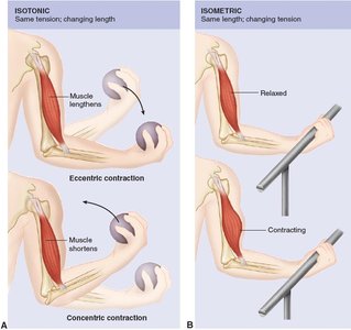
Isotonic contraction: Muscle changes length, producing movement (includes concentric and eccentric contractions).
Isometric contraction: Muscle tension increases without changing length; no movement occurs.
Muscle Roles in Movement
Prime mover: Main muscle responsible for a movement.
Synergist: Assists the prime mover.
Antagonist: Opposes the action of the prime mover.
Posture and Heat Production
Tonic contractions help maintain posture by counteracting gravity. Muscle contractions also generate heat, which is essential for maintaining body temperature.
Fatigue and Oxygen Debt
Muscle fatigue results from prolonged activity, leading to ATP depletion and lactic acid accumulation. Oxygen debt is the extra oxygen required to metabolize lactic acid after exercise.
Integration with Other Body Systems
Muscle function depends on the nervous, skeletal, respiratory, and circulatory systems. Disorders in these systems can impair movement, as seen in conditions like multiple sclerosis or spinal cord injury.
Neuromuscular Junction and Motor Units
Motor Unit Structure and Function
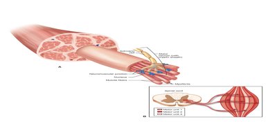
A motor unit consists of a motor neuron and the muscle fibers it innervates. The neuromuscular junction (NMJ) is where the nerve meets the muscle fiber, and acetylcholine (ACh) is the neurotransmitter that triggers muscle contraction.
Muscle Stimulus and Response
Threshold stimulus: Minimum stimulus required for muscle contraction.
All-or-none response: Individual muscle fibers contract fully or not at all when stimulated above threshold.
Graded response: Whole muscles can produce varying degrees of force by recruiting different numbers of motor units.
Effects of Exercise on Muscles
Muscle Adaptation
Regular exercise improves muscle tone, posture, and endurance. Disuse leads to atrophy, while strength training causes hypertrophy (increase in muscle size). Endurance training enhances the muscle's ability to sustain activity but does not significantly increase size.
Movements Produced by Skeletal Muscles
Types of Movements
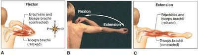
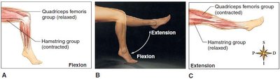
Flexion: Decreases the angle between bones (e.g., bending the elbow).
Extension: Increases the angle between bones (e.g., straightening the knee).
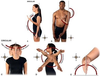
Abduction: Moves a limb away from the midline.
Adduction: Moves a limb toward the midline.
Rotation: Movement around a longitudinal axis.
Circumduction: Circular movement of a limb.
Supination/Pronation: Rotational movements of the forearm.
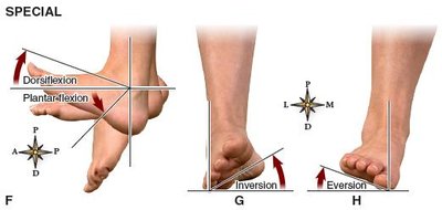
Dorsiflexion/Plantar flexion: Movements of the foot upward or downward.
Inversion/Eversion: Turning the sole of the foot inward or outward.
Muscle Groups and Actions
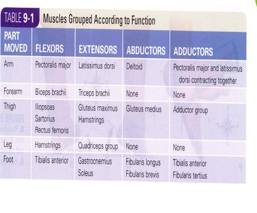
Muscles are grouped by their actions at joints. The following table summarizes major muscle groups and their functions:
Part Moved | Flexors | Extensors | Abductors | Adductors |
|---|---|---|---|---|
Arm | Pectoralis major | Latissimus dorsi | Deltoid | Pectoralis major and latissimus dorsi contracting together |
Forearm | Biceps brachii | Triceps brachii | None | None |
Thigh | Iliopsoas, Sartorius, Rectus femoris | Gluteus maximus, Hamstrings | Gluteus medius | Adductor group |
Leg | Hamstrings | Quadriceps group | None | None |
Foot | Tibialis anterior | Gastrocnemius, Soleus | Fibularis longus, Fibularis brevis | Tibialis anterior, Fibularis tertius |
Major Skeletal Muscle Groups
Muscles of the Head and Neck
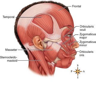
Facial muscles: Orbicularis oculi, orbicularis oris, zygomaticus (smiling muscle).
Muscles of mastication: Masseter and temporal (close the jaw).
Sternocleidomastoid: Flexes the head.
Trapezius: Elevates shoulders and extends the head.
Muscles of the Upper Extremities
Pectoralis major: Flexes upper arm.
Latissimus dorsi: Extends upper arm.
Deltoid: Abducts upper arm.
Biceps brachii: Flexes forearm.
Triceps brachii: Extends forearm.
Muscles of the Trunk
Abdominal muscles: Rectus abdominis, external oblique, internal oblique, transversus abdominis.
Respiratory muscles: Intercostal muscles, diaphragm.
Muscles of the Lower Extremities
Iliopsoas: Flexes thigh.
Gluteus maximus: Extends thigh.
Adductor muscles: Adduct thighs.
Hamstring muscles: Flex leg (semimembranosus, semitendinosus, biceps femoris).
Quadriceps femoris group: Extend leg (rectus femoris, vastus muscles).
Tibialis anterior: Dorsiflexes foot.
Gastrocnemius: Plantar flexes foot.
Fibularis group: Flexes foot.
Muscular Disorders
Muscle Injuries and Infections
Strain: Overexertion or trauma causing muscle fiber damage.
Cramps: Painful, involuntary muscle contractions.
Contusion: Muscle bruise from trauma.
Crush injury: Severe trauma releasing myoglobin, potentially causing kidney failure.
Poliomyelitis: Viral infection of motor nerves, may cause paralysis.
Tetanus: Caused by Clostridium tetani toxin, leads to sustained muscle contractions.
Muscular Dystrophy
Duchenne muscular dystrophy (DMD): X-linked genetic disorder causing muscle wasting, primarily in boys.
Characterized by progressive weakness, pseudohypertrophy, and early onset.
Myasthenia Gravis
Autoimmune disease causing muscle weakness, especially in the face and throat.
Results from immune attack on ACh receptors at the NMJ.
Treatment may include ACh breakdown inhibitors and immunosuppressants.
Clinical and Applied Aspects
Rigor Mortis
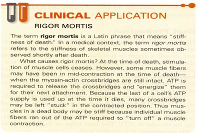
Rigor mortis is the postmortem stiffening of muscles due to the depletion of ATP, preventing the release of myosin-actin cross-bridges.
Muscle Fiber Types
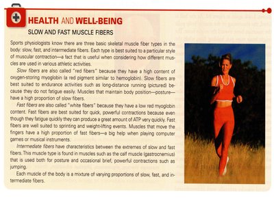
There are three main types of skeletal muscle fibers: slow (red), fast (white), and intermediate. Slow fibers are fatigue-resistant and suited for endurance, while fast fibers contract quickly but fatigue rapidly. Intermediate fibers have properties between the two.
Enhancing Muscle Strength
Strength training increases muscle size and strength. Chemical enhancements, such as anabolic steroids, can increase muscle mass but carry significant health risks. Proper nutrition and training are essential for safe muscle development.
Muscle Massage
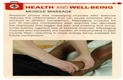
Massage after exercise can reduce inflammation, soreness, and increase the number of mitochondria in muscle fibers, enhancing recovery and future performance.
Review Questions
A muscle’s attachment to a stationary bone is called its origin.
The two proteins composing myofilaments are actin and myosin.
The primary functions of muscles are movement, posture (muscle tone), and heat production.
The point of contact between a nerve ending and the muscle fiber is called the neuromuscular junction.
When the muscle contracts and no movement results, it is called an isometric contraction.
The muscle that functions in closing the jaw is the masseter.
Abduction means moving a part away from the midline of the body.
The muscular disease characterized by the immune system attacking muscle cells at the neuromuscular junction is myasthenia gravis.
