 Back
The Muscular System: Structure, Function, and Contraction
Study Guide - Smart Notes
 Tailored notes based on your materials, expanded with key definitions, examples, and context.
The Muscular System
Types of Muscle Tissue
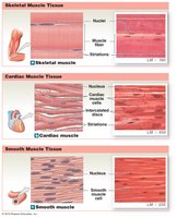
The human body contains three distinct types of muscle tissue, each with unique structural and functional characteristics:
Skeletal Muscle Tissue: Cylindrical, striated, multinucleate cells with nuclei at the periphery. Found attached to the skeletal system and under voluntary control.
Cardiac Muscle Tissue: Branched, striated cells with one or two central nuclei and intercalated discs. Located only in the heart and under involuntary control.
Smooth Muscle Tissue: Spindle-shaped, non-striated cells with a single central nucleus. Found in the walls of hollow organs (e.g., GI tract, blood vessels) and under involuntary control.
Functions of Skeletal Muscle
Skeletal muscles perform several essential functions in the body:
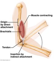
Produce Movement: Muscles pull on bones to create lever-like movements.
Maintain Posture: Enable the body to remain upright while sitting or standing.
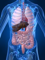
Support Soft Tissues: Abdominal muscles protect and support internal organs.
Guard Entrances and Exits: Muscles encircle openings of digestive and urinary tracts, allowing voluntary control.
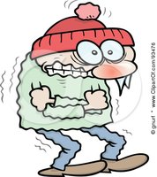
Maintain Body Temperature: Muscle contractions generate heat, especially during shivering, helping regulate body temperature.
Gross Anatomy of Skeletal Muscle
Muscle as an Organ
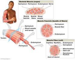
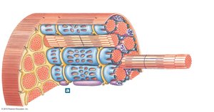
Each skeletal muscle is an organ composed of muscle tissue, connective tissue, blood vessels, and nerves. Muscle cells are called muscle fibers due to their elongated shape.
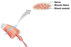
Levels of Organization
Skeletal muscle is organized into hierarchical structures:
Muscle: The entire organ.
Muscle Fascicle: Bundles of muscle fibers.
Muscle Fiber: Individual muscle cells.
Connective Tissue Layers
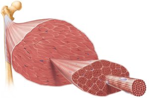
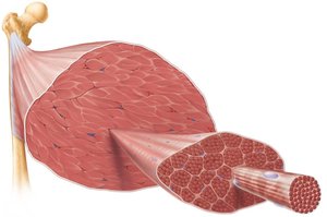
Three layers of connective tissue organize and protect skeletal muscle:
Epimysium: Surrounds the entire muscle (dense irregular connective tissue).
Perimysium: Surrounds each fascicle (fibrous connective tissue).
Endomysium: Surrounds each muscle fiber (loose connective tissue with reticular fibers).
Layer | Location | Function |
|---|---|---|
Epimysium | Surrounds entire muscle | Protects and binds muscle |
Perimysium | Surrounds fascicles | Bundles fibers, supports blood vessels/nerves |
Endomysium | Surrounds muscle fibers | Supports individual fibers |
Microscopic Anatomy of Skeletal Muscle Fiber
Unique Features of Muscle Fibers
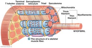
Skeletal muscle fibers are large, multinucleate cells with nuclei at the periphery. They contain specialized structures:
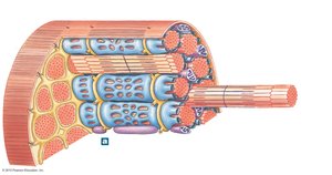
Sarcolemma: Plasma membrane of the muscle fiber.
Sarcoplasm: Cytoplasm of the muscle fiber.
Sarcoplasmic Reticulum (SR): Smooth endoplasmic reticulum specialized for calcium storage, crucial for muscle contraction.
T-Tubules, Terminal Cisternae, and Triads
Muscle fibers contain additional specialized structures:
T-Tubules: Transverse tubules that conduct electrical signals deep into the fiber.
Terminal Cisternae: Enlarged regions of the SR adjacent to T-tubules.
Triad: Structure formed by a T-tubule flanked by two terminal cisternae.
Myofibrils and Myofilaments
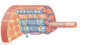
Myofibrils are cylindrical organelles responsible for contraction, making up ~80% of muscle fiber volume. They are composed of myofilaments:
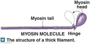
Thick Filaments: Made of myosin.
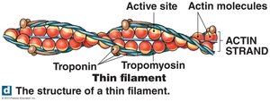
Thin Filaments: Made of actin, troponin, and tropomyosin.
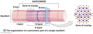
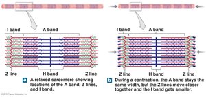
Myofilaments are organized into sarcomeres, the functional unit of contraction, defined as the segment between two Z-discs.
Region | Contents |
|---|---|
Z line | Boundary of sarcomere |
I band | Thin filaments only |
A band | Thick filaments, some overlap with thin |
H band | Thick filaments only |
M line | Proteins connecting thick filaments |
Structure of Myofilaments
Thin filaments consist of actin molecules with active sites, covered by tropomyosin and regulated by troponin. Thick filaments are composed of myosin molecules with heads that bind to actin during contraction.
Muscle Contraction
Contraction of the Sarcomere
Muscle contraction begins with the contraction of the sarcomere. The process involves:
Increase in intracellular calcium as Ca2+ is released from the sarcoplasmic reticulum.
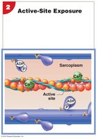
Ca2+ binds to troponin, causing tropomyosin to shift and expose actin's active site.
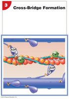
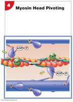
Myosin binds to actin, pivots toward the center of the sarcomere, and pulls the thin filament, shortening the sarcomere.
Neuromuscular Junction (NMJ)
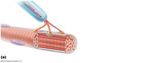
Skeletal muscle contraction is initiated by stimulation from a motor neuron at the neuromuscular junction:
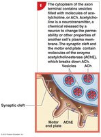
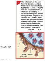
Axon Terminal: Contains synaptic vesicles with acetylcholine (ACh).
Motor End Plate: Specialized region of sarcolemma with ACh receptors.
Synaptic Cleft: Space between axon terminal and motor end plate.
Acetylcholinesterase (AChE): Enzyme that breaks down ACh.
Steps at the Neuromuscular Junction
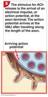
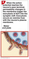
Action potential travels down the axon to the synaptic end bulb, opening voltage-gated calcium channels.
Calcium influx triggers exocytosis of ACh-containing vesicles.
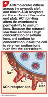
ACh is released into the synaptic cleft and binds to receptors, opening ligand-gated ion channels and allowing sodium influx.
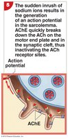
Sodium influx depolarizes the motor end plate, triggering a muscle action potential.
ACh is degraded by AChE, ending the action potential.
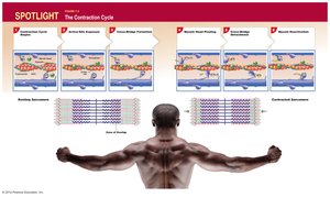
Contraction Cycle
The contraction cycle consists of several steps:
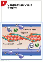
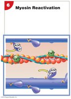
Myosin head is "cocked" by ATP hydrolysis; Ca2+ binds to troponin, shifting tropomyosin and exposing actin's active site.
Cross-bridge forms as myosin binds to actin.
Myosin head pivots (powerstroke), pulling the thin filament and shortening the sarcomere.
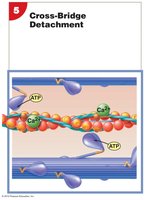
Detachment occurs when new ATP binds to myosin, causing it to release from actin.
Cycle repeats as long as ATP and Ca2+ are present.
Motor Units
Definition and Function
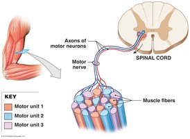
A motor unit consists of a single motor neuron and all the skeletal muscle fibers it innervates. Each muscle fiber is innervated by only one motor neuron. Motor units allow for precise control of muscle contraction.
Summary Table: Muscle Contraction Steps
Step | Event | Key Molecules |
|---|---|---|
1 | AP arrives at NMJ | ACh, Ca2+ |
2 | ACh released, binds to receptors | ACh |
3 | Sodium influx, muscle AP | Na+ |
4 | Ca2+ released from SR | Ca2+ |
5 | Cross-bridge formation | Myosin, Actin |
6 | Powerstroke | ATP, Myosin |
7 | Detachment and reactivation | ATP |
Key Equations
ATP hydrolysis provides energy for muscle contraction:
Calcium release and binding:
Additional info: The notes have been expanded with academic context, including definitions, examples, and tables for clarity. All images included are directly relevant to the adjacent content and reinforce anatomical and physiological concepts.
