 Back
The Muscular System: Structure, Function, and Physiology
Study Guide - Smart Notes
 Tailored notes based on your materials, expanded with key definitions, examples, and context.
Chapter 9: The Muscular System
Overview
This chapter provides a comprehensive overview of the muscular system, including muscle types, structure, function, physiology of contraction, energy sources, and clinical considerations. The muscular system is essential for movement, posture, joint stabilization, temperature regulation, and the control of substances within the body.
Functions of Muscle
Main Functions
Movement: Muscles contract to produce movement of the body and its parts.
Maintaining Posture: Continuous muscle contractions maintain body posture.
Stabilizing Joints: Muscles reinforce and stabilize joints.
Temperature Homeostasis: Muscle activity generates heat, helping maintain body temperature.
Regulation of Substances: Muscles control the movement of substances between body compartments (e.g., sphincters).
Characteristics of Muscle Tissue
Key Properties
Excitability: Ability to receive and respond to stimuli.
Contractility: Ability to shorten forcibly when stimulated.
Extensibility: Ability to be stretched beyond resting length.
Elasticity: Ability to return to original length after stretching.
Muscle Types
Classification and Features
Skeletal Muscle: Attached to bones, voluntary, striated, limited regeneration.
Cardiac Muscle: Found in heart walls, involuntary, striated, no regeneration.
Smooth Muscle: Located in walls of hollow organs, involuntary, non-striated, fast regeneration.
Skeletal Muscle Structure
Connective Tissue Sheaths
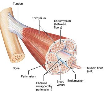
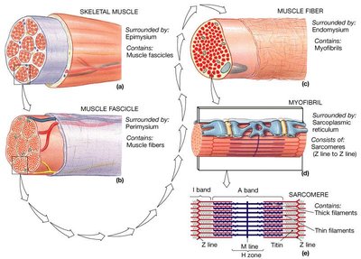
Epimysium: Dense connective tissue surrounding the whole muscle.
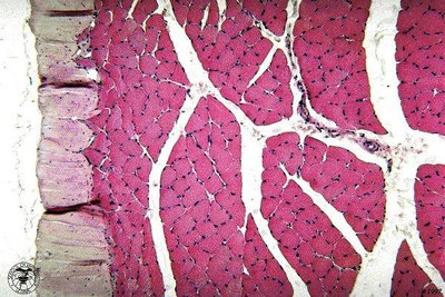
Perimysium: Fibrous connective tissue surrounding fascicles (bundles of muscle fibers).
Endomysium: Fine areolar connective tissue surrounding each muscle fiber.
Muscle Attachments
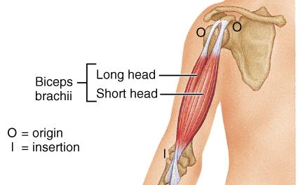
Origin: Attachment to bone that does not move during contraction.
Insertion: Attachment to bone that moves during contraction.
Direct Attachment: Epimysium fused to periosteum or perichondrium.
Indirect Attachment: Connective tissue extends as tendon or aponeurosis; more common due to durability and size.
Microscopic Anatomy of Skeletal Muscle
Muscle Fiber Structure
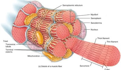
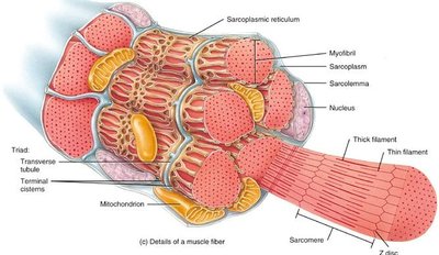
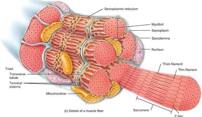
Muscle Fiber: Long, cylindrical, multinucleated cell.
Sarcolemma: Muscle cell membrane; forms T-tubules for rapid signal transmission.
Sarcoplasm: Cytoplasm containing glycogen, myoglobin, and creatine phosphate for energy.
Nucleus: Multiple nuclei pushed to the side due to myoblast fusion.
Mitochondria: Numerous for energy production.
Sarcoplasmic Reticulum (SR): Specialized smooth ER for Ca2+ storage.
Triad: Structure formed by T-tubule and two terminal cisterns of SR, crucial for excitation-contraction coupling.
Myofibrils and Sarcomeres
Myofibrils: Rod-like structures containing contractile proteins.
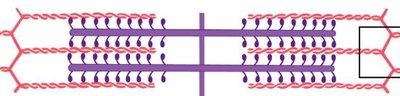
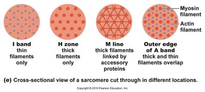
Sarcomere: Functional contractile unit formed by thick and thin filaments.
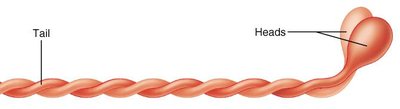
Thick Filaments: Composed of myosin molecules; each has a head (cross bridge) with actin and ATP binding sites.
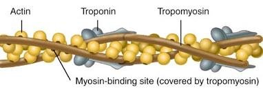
Thin Filaments: Composed of actin, tropomyosin, and troponin; actin has myosin binding sites.
Muscle Physiology: Contraction
Ion Channels and Membrane Potential
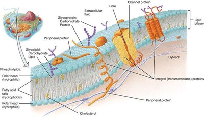
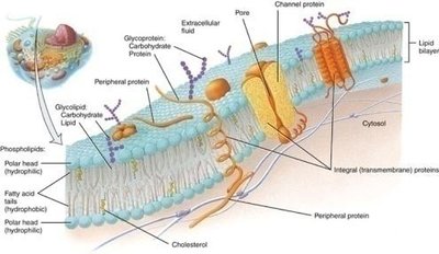
Ion Channels: Allow ions to diffuse across the cell membrane.
Gated Channels: Open in response to voltage changes (voltage-gated) or ligand binding (ligand-gated).
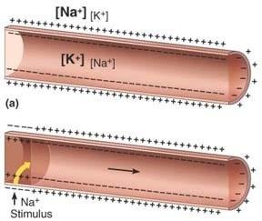
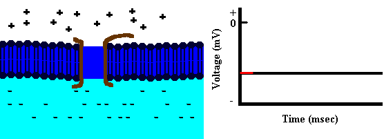
Resting Membrane Potential: Maintained by Na+/K+ pump; typically -90 mV in muscle cells.
Steps of Muscle Contraction
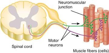
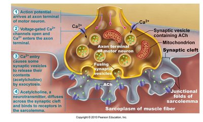
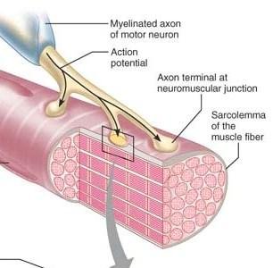
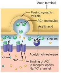
Excitation: Nerve impulse triggers release of acetylcholine (ACh) at neuromuscular junction.
Excitation-Contraction Coupling: Action potential travels along sarcolemma and T-tubules, causing Ca2+ release from SR.
Contraction: Ca2+ binds to troponin, moving tropomyosin and exposing myosin binding sites on actin; myosin heads bind and pull actin (power stroke).
Relaxation: Ca2+ is reabsorbed into SR, tropomyosin covers binding sites, and muscle returns to resting state.
Sliding Filament Theory
ATP Hydrolysis: Myosin head hydrolyzes ATP, energizing for binding.
Crossbridge Formation: Myosin head binds to actin, releasing phosphate.
Power Stroke: Myosin head pivots, pulling actin toward sarcomere center.
Detachment: ATP binds to myosin, causing detachment from actin.
Cycle Repeats: As long as Ca2+ and ATP are available.
Equation for ATP hydrolysis:
Motor Units and Muscle Contraction
Motor Units
Motor Unit: A motor neuron and all the muscle fibers it innervates.
Size: Small motor units allow fine control; large units provide powerful contractions.
All-or-None Principle: Each muscle fiber contracts fully when stimulated.
Graded Response: Muscle contraction strength depends on number and frequency of motor units activated.
Types of Muscle Contraction
Isotonic: Muscle changes length, moves load (concentric: shortens; eccentric: lengthens).
Isometric: Tension increases, but muscle length does not change.
Smooth Muscle Contraction
Gross and Microscopic Anatomy
Location: Walls of hollow organs, arranged in layers.
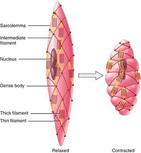
Cell Structure: Small, spindle-shaped, single nucleus, no T-tubules, gap junctions, few mitochondria.
Myofibrils: Actin and myosin present, no sarcomeres, intermediate filaments anchor actin.
Regulatory Proteins: Tropomyosin present, calmodulin replaces troponin.
Physiology of Smooth Muscle Contraction
Stimulation: Can be autonomic, hormonal, or local.
Diffuse Junction: Neurotransmitter released from varicosities into wide cleft, affecting many cells.
Contraction: Slow, synchronized, resistant to fatigue, uses little energy.
Energy for Muscle Contraction
ATP and Regeneration Mechanisms
ATP: Direct energy source for crossbridge cycling; only enough stored for 4-6 seconds.
Creatine Phosphate: Regenerates ATP for short bursts (15 seconds).
Anaerobic Respiration (Glycolysis): Rapid ATP production, produces lactic acid, fuels muscles for up to 45 seconds.
Aerobic Respiration: Requires O2, produces large amounts of ATP, supports prolonged activity.
Equation for ATP regeneration:
Muscle Cell Types
Slow Oxidative: Thin, slow ATPases, abundant myoglobin and mitochondria, aerobic, endurance, not powerful.
Fast Glycolytic: Large diameter, fast ATPases, little myoglobin, few mitochondria, anaerobic, powerful, fatigues quickly.
Fast Oxidative: Intermediate size, fast ATPases, abundant myoglobin, uses both aerobic and anaerobic, fatigue resistant, intermediate power.
Effect of Exercise on Muscles
Aerobic Exercise: Increases capillaries, mitochondria, and myoglobin; converts fast glycolytic to fast oxidative.
Resistance Exercise: Increases muscle cell size, myofibrils, and glycogen stores; converts fast oxidative to fast glycolytic.
Clinical Considerations
Muscular Dystrophy
Duchenne Muscular Dystrophy (DMD): X-linked recessive disorder; lack of dystrophin protein destabilizes sarcolemma, leading to muscle degeneration and early death.
Rigor Mortis
Occurs after death due to Ca2+ influx and lack of ATP, causing myosin heads to remain attached to actin, resulting in muscle rigidity.
Disappears as proteolytic enzymes degrade myofilaments.
Review Questions
What are the three muscle membranes?
What is the structure of skeletal muscle from the outside in?
What is another name for a muscle cell?
Name the protein filaments of the sarcomere.
What is the sarcomere?
Name the three proteins that make up actin. Which one binds calcium?
What is the trigger for ACh release? Where does it happen?
What is an action potential?
Where is Ca released from in the muscle cell?
What is a cross bridge?
What happens when Ca levels in the muscle cell drop?
