 Back
The Muscular System: Structure, Function, and Major Muscles
Study Guide - Smart Notes
 Tailored notes based on your materials, expanded with key definitions, examples, and context.
The Muscular System
Structure of Skeletal Muscles
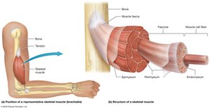
Skeletal muscles are complex organs composed of muscle fibers, connective tissues, blood vessels, and nerves. Their organization allows for efficient force generation and movement.
Skeletal Muscle Fibers: Long, cylindrical cells surrounded by a thin layer of extracellular matrix called the endomysium.
Fascicle: A bundle of 10–100 muscle fibers, surrounded by perimysium.
Epimysium: Connective tissue sheath that surrounds all fascicles in a muscle.
Fascia: The most superficial connective tissue sheath, continuous with the epimysium.
Tendons: Attach muscle to bone or other structures, transmitting the force of contraction.
Vascular and Neural Supply: Skeletal muscles are richly supplied with blood vessels and nerves, essential for function and repair.
Voluntary Control: Skeletal muscle contraction is under conscious control and requires nervous stimulation.
Muscle Knots (Myofascial Trigger Points)
Muscle knots are localized, irritable spots within the fascia surrounding a muscle, often resulting in pain and restricted movement.
Causes: Repetitive exercise, trauma, stress, disease, or trapped nerves can lead to trigger points.
Symptoms: Sustained contraction and pain at the site.
Treatment: Includes massage, anti-inflammatory medications, muscle relaxants, and passive stretching.
Fascicle Patterns and Muscle Shapes
The arrangement of fascicles within a muscle influences its range of motion and power.
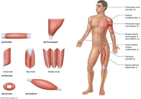
Parallel: Fascicles run parallel to the muscle's long axis (e.g., Sartorius).
Convergent: Broad origin, fascicles converge to a single tendon (e.g., Pectoralis major).
Circular (Sphincter): Fascicles arranged in concentric rings (e.g., Orbicularis oculi).
Fusiform: Thick in the middle, tapered at ends (e.g., Biceps brachii).
Pennate: Fascicles attach obliquely to a tendon (resembles a feather):
Unipennate: Fascicles on one side (e.g., Flexor pollicis longus).
Bipennate: Fascicles on both sides (e.g., Rectus femoris).
Multipennate: Multiple tendons (e.g., Deltoid).
Naming Muscles
Muscles are named based on several criteria, reflecting their characteristics or functions.
Size: Major, minor, longus, brevis, vastus.
Location: Directional (superior, inferior, medial, lateral) or regional terms (e.g., Pectoralis minor).
Attachment: Named for bones or structures attached (e.g., Sternocleidomastoid).
Function: Flexor, extensor, adductor, abductor, levator.
Depth: Profundus (deep), superficialis (superficial).
Functions of Skeletal Muscles
Skeletal muscles contract to generate force, enabling movement and other vital functions.
Muscle Tension: The force generated by muscle contraction.
Body Movements: Muscles produce actions by moving bones at joints.
Heat Generation: Conversion of ATP to mechanical energy produces heat (e.g., shivering).
Functional Groups of Muscles
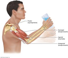
Agonist (Prime Mover): Main muscle responsible for movement.
Antagonist: Opposes or slows the agonist's action.
Synergist: Assists the agonist, stabilizing movement.
Fixator: Stabilizes the origin of the agonist for efficient movement.
Muscle Origin and Insertion
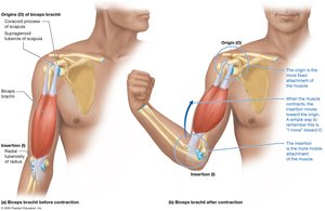
Origin (O): The fixed attachment point of a muscle.
Insertion (I): The moveable attachment point.
Example: Biceps brachii originates on the scapula and inserts on the radius.
Origin and insertion can switch depending on the movement (e.g., Iliopsoas).
Lever Systems in Body Movements
Muscles, bones, and joints form lever systems to move loads efficiently.
Lever: Bone that moves on a fixed point (fulcrum).
Load: Object or body part being moved.
Force: Muscle tension applied to the lever.
Fulcrum: Joint acting as the pivot point.
Types of Levers
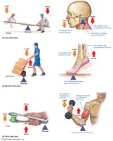
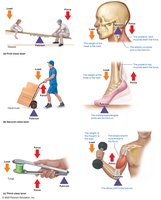
First-Class Lever: Fulcrum between load and force (e.g., atlanto-occipital joint).
Second-Class Lever: Load between fulcrum and force (e.g., metatarsophalangeal joints; uncommon in body).
Third-Class Lever: Force between load and fulcrum (e.g., elbow joint; most common in body).
Mechanical Advantage and Disadvantage
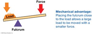
Mechanical Advantage: Small force moves a large load (fulcrum close to load).
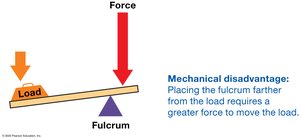
Mechanical Disadvantage: Greater force required to move the load (fulcrum farther from load).
Muscle Strains
Muscle strains occur when a muscle is overstretched or subjected to excessive force, resulting in tears.
Symptoms: Pain, limited movement, swelling, bruising.
Treatment: PRICE method (Protect, Rest, Ice, Compression, Elevation), medications, physical therapy, or surgery.
Studying Muscles: Regional Overview
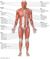
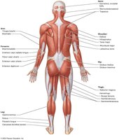
Muscles are categorized by region for systematic study:
Head, neck, and vertebral column
Trunk and pelvic floor
Pectoral girdle and upper limb
Hip and lower limb
Concept Boost: Demystifying Muscle Actions
Determining Muscle Actions
Muscle actions can be deduced by analyzing their origins and insertions. The muscle typically moves the insertion toward the origin during contraction.
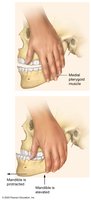
Fingertip Method: Place the base of your palm over the origin and fingertips over the insertion; mimic contraction by pulling fingertips toward the palm.
Example: Medial pterygoid muscle pulls the mandible up and forward.
Muscles of Facial Expression
Overview
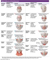
Facial muscles insert into skin or connective tissue, allowing for a wide range of expressions.
Epicranius: Elevates eyebrows and forehead skin.
Orbicularis oculi: Closes eyelids (blinking, winking).
Levator palpebrae superioris: Opens eyelids.
Zygomaticus major/minor: Smiling.
Risorius: Lateral movement of mouth corners (smirk).
Orbicularis oris: Controls lips for speech, eating, whistling.
Depressor anguli oris, depressor labii inferioris, mentalis: Frowning, pouting.
Buccinator: Sucking, chewing, whistling.
Platysma: Jaw-dropping, tightens neck skin.
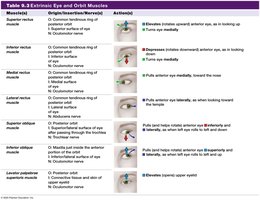
Extrinsic Eye Muscles
Overview
Six extrinsic muscles control eye movement, originating from the common tendinous ring.
Medial rectus: Moves eye medially.
Lateral rectus: Moves eye laterally.
Superior rectus: Moves eye superiorly and medially.
Inferior rectus: Moves eye inferiorly and medially.
Superior oblique: Rotates eye inferiorly and laterally (passes through trochlea).
Inferior oblique: Rotates eye superiorly and laterally.
Muscles of Mastication
Overview
Muscles of mastication move the mandible for chewing.
Masseter: Elevates mandible.
Temporalis: Elevates mandible.
Medial pterygoid: Elevates mandible, assists with side-to-side movement.
Lateral pterygoid: Depresses and protracts mandible, side-to-side movement.
Muscles That Move the Head and Neck
Overview
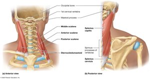
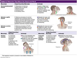
Sternocleidomastoid: Rotates head to opposite shoulder, flexes head when both sides contract.
Scalene muscles (anterior, middle, posterior): Lateral flexion of head, elevate ribs during deep breathing.
Trapezius (superior part): Extends head.
Splenius capitis/cervicis: Rotate and extend head and neck.
Muscles of the Vertebral Column
Erector Spinae Muscle Group
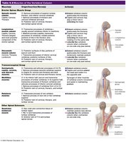
Major postural muscles that extend and laterally flex the vertebral column.
Lateral column: Iliocostalis lumborum, thoracis, cervicis.
Middle column: Longissimus thoracis, cervicis, capitis.
Medial column: Spinalis thoracis, cervicis, capitis.
Transversospinal Group
Semispinalis, multifidus, rotatores: Extend and rotate vertebral column.
Quadratus lumborum: Extends and laterally flexes vertebral column.
Muscles of Ventilation
Overview
Muscles involved in breathing alter the size of the thoracic cavity.
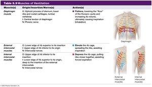
Diaphragm: Main muscle of inspiration; contracts and flattens to increase thoracic volume.
External intercostals: Elevate and expand rib cage (inspiration).
Internal intercostals: Depress rib cage (forced expiration).
Sternocleidomastoid and scalenes: Assist in forced inspiration.
Abdominal Muscles
Overview
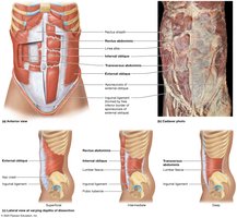
Abdominal muscles support trunk movement and increase intra-abdominal pressure.
Rectus abdominis: Flexes trunk, enclosed by rectus sheath, divided by linea alba.
External and internal obliques: Rotate and laterally flex trunk.
Transversus abdominis: Compresses abdominal cavity.
Muscles That Move the Scapula at the Pectoral Girdle
Overview
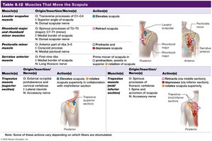
Serratus anterior: Protracts and rotates scapula superiorly.
Pectoralis minor: Protracts and depresses scapula.
Trapezius: Superior part elevates, middle part retracts, inferior part depresses scapula.
Levator scapulae: Elevates scapula.
Rhomboid major/minor: Retract scapula.
Muscles That Move the Arm at the Shoulder Joint
Overview
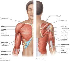
Pectoralis major: Flexes, adducts, and internally rotates arm.
Coracobrachialis: Assists with flexion and adduction of humerus.
Deltoid: Anterior fibers flex, central fibers abduct, posterior fibers extend arm.
Latissimus dorsi: Extends, adducts, and internally rotates arm.
Teres major: Assists latissimus dorsi.
Rotator cuff muscles: Stabilize shoulder joint (supraspinatus, infraspinatus, teres minor, subscapularis).
Muscles That Move the Forearm and Hand
Overview
Biceps brachii: Flexes and supinates forearm.
Brachialis: Agonist of elbow flexion.
Brachioradialis: Assists elbow flexion.
Triceps brachii: Extends forearm.
Anconeus: Assists triceps brachii.
Flexors (anterior/medial forearm): Flex hand and fingers.
Extensors (posterior/lateral forearm): Extend hand and fingers.
Muscles of the Hip and Lower Limb
Hip Muscles
Iliopsoas: Main thigh flexor.
Pectineus: Assists thigh flexion and adduction.
Adductor group (magnus, longus, brevis): Adduct thigh.
Gracilis: Adducts thigh.
Sartorius: Flexes, abducts, laterally rotates thigh; flexes leg.
Gluteal group: Gluteus maximus (extends, abducts, laterally rotates thigh), medius and minimus (abduct, medially rotate thigh).
Lateral rotators: Piriformis, obturator internus/externus, gemelli, quadratus femoris.
Knee Muscles
Quadriceps femoris: Four muscles (rectus femoris, vastus lateralis, intermedius, medialis) extend leg at knee; rectus femoris also flexes thigh.
Hamstrings: Semitendinosus, semimembranosus, biceps femoris; extend thigh and flex leg.
Ankle, Foot, and Toe Muscles
Tibialis anterior, extensor digitorum longus: Dorsiflex foot; tibialis anterior inverts foot.
Extensor hallucis longus: Extends big toe, assists dorsiflexion.
Fibularis longus/brevis: Evert foot.
Gastrocnemius, soleus (triceps surae): Plantarflex foot; gastrocnemius also flexes leg.
Plantaris, tibialis posterior: Plantarflex foot; tibialis posterior assists inversion.
Common Injuries
Rotator cuff injuries: Overuse or trauma can cause tears, especially in the supraspinatus tendon; symptoms include pain, weakness, and limited range of motion.
Calcaneal tendon injuries: Common in athletes; may result in pain, swelling, and loss of plantarflexion; diagnosed with the Simmonds test.
The Big Picture of Muscle Movement
Muscle contraction, coordinated by the nervous system, enables all voluntary movements, maintains posture, and generates heat, highlighting the essential role of the muscular system in human physiology.
