Back
BackThe Muscular System: Structure, Function, and Mechanisms
Study Guide - Smart Notes
 Tailored notes based on your materials, expanded with key definitions, examples, and context.
The Muscular System
Overview of Muscle Types
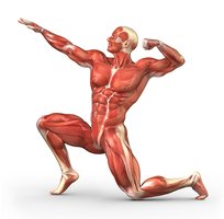
Muscles are essential for all types of body movement and are classified into three basic types: skeletal muscle, cardiac muscle, and smooth muscle. Each type has distinct structural and functional characteristics.
Skeletal muscle: Responsible for voluntary movements, attached to bones.
Cardiac muscle: Found only in the heart, responsible for pumping blood.
Smooth muscle: Located in walls of hollow organs, controls involuntary movements.
Comparison of Muscle Types
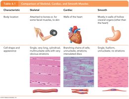
Characteristic | Skeletal | Cardiac | Smooth |
|---|---|---|---|
Body location | Attached to bones or skin | Walls of the heart | Walls of hollow organs |
Cell shape & appearance | Long, cylindrical, multinucleate, striated | Branching, single nucleus, striated, intercalated discs | Spindle-shaped, single nucleus, no striations |
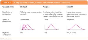
Regulation of contraction | Voluntary (nervous system) | Involuntary (pacemaker, nervous system, hormones) | Involuntary (nervous system, hormones, chemicals, stretch) |
Speed of contraction | Slow to fast | Slow | Very slow |
Rhythmic contraction | No | Yes | Yes, in some |
Skeletal Muscle Structure
Connective Tissue Wrappings
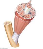
Skeletal muscle fibers are organized and protected by several layers of connective tissue:
Endomysium: Surrounds each individual muscle fiber.
Perimysium: Wraps around bundles of fibers called fascicles.
Epimysium: Encloses the entire muscle.
Fascia: Outermost layer, separates muscles from surrounding tissues.
Skeletal Muscle Attachments
Muscles attach to bones via tendons, which are tough, cordlike structures composed mainly of collagen. Tendons often cross joints, allowing for efficient transfer of force.
Smooth and Cardiac Muscle Structure
Smooth Muscle
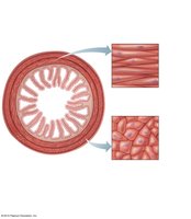
Smooth muscle cells are spindle-shaped, have a single nucleus, and lack striations. They are found in the walls of hollow organs such as the stomach, bladder, and blood vessels, and are controlled involuntarily. 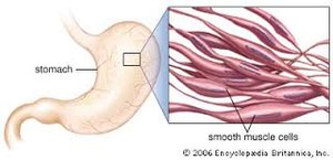
Cardiac Muscle
Cardiac muscle cells are striated, branching, and usually have a single nucleus. They are joined by intercalated discs and are found only in the heart, where they contract involuntarily. 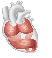
Functions of Skeletal Muscle
Major Functions
Skeletal muscles perform several vital functions:
Produce movement: Facilitate locomotion and manipulation.
Maintain posture: Stabilize body position.
Stabilize joints: Support and reinforce joints.
Generate heat: Maintain body temperature through contraction.
Microscopic Anatomy of Skeletal Muscle
Muscle Fiber Structure
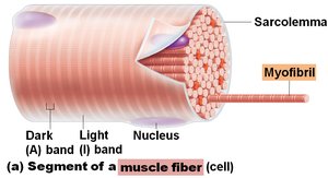
Each muscle fiber (cell) contains:
Sarcolemma: Specialized plasma membrane.
Myofibrils: Long organelles responsible for contraction, composed of repeating units called sarcomeres.
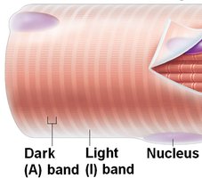
Banding pattern: Alternating light (I) and dark (A) bands create striations.
Sarcomere Organization
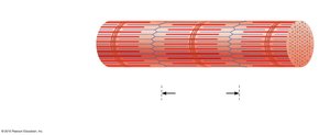
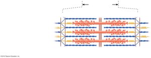
The sarcomere is the contractile unit of muscle, defined by Z discs. It contains:
Thick filaments: Composed of myosin, with heads forming cross-bridges during contraction.
Thin filaments: Composed of actin, anchored to Z discs.
H zone: Central area lacking actin filaments when muscle is relaxed.
M line: Center of the H zone.
Sarcoplasmic Reticulum
The sarcoplasmic reticulum (SR) is a specialized smooth endoplasmic reticulum that stores and releases calcium ions, essential for muscle contraction.
Muscle Cell Properties
Functional Characteristics
Muscle cells exhibit several key properties:
Irritability (Responsiveness): Ability to respond to stimuli.
Contractility: Ability to shorten when stimulated.
Extensibility: Ability to be stretched.
Elasticity: Ability to return to original length after stretching.
Stimulation and Contraction
Nerve Stimulus and Action Potential
Skeletal muscles contract only when stimulated by a motor neuron. The motor unit consists of one motor neuron and all the muscle fibers it stimulates. 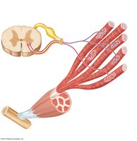
Neuromuscular Junction
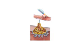
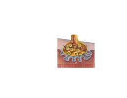
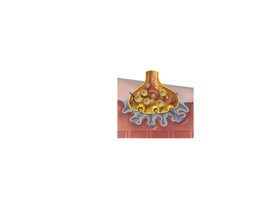
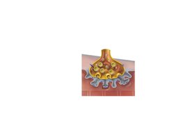
The neuromuscular junction is the site where the axon terminal of a motor neuron meets the sarcolemma of a muscle fiber. The neurotransmitter acetylcholine (ACh) is released upon nerve impulse arrival, triggering muscle contraction.
Transmission of Nerve Impulse
Calcium channels open in the axon terminal, allowing Ca2+ entry.
Ca2+ causes synaptic vesicles to release ACh by exocytosis.
ACh diffuses across the synaptic cleft and binds to receptors on the sarcolemma.
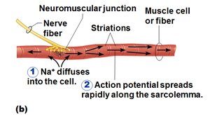
Sodium (Na+) enters the cell, potassium (K+) leaves, causing depolarization and action potential.
Acetylcholinesterase (AChE) breaks down ACh, ending contraction.
Action Potential Analogy
The spread of an action potential along the sarcolemma is analogous to a flame rapidly consuming a dry twig. 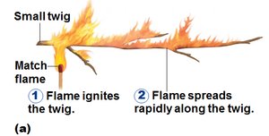
Mechanism of Muscle Contraction: Sliding Filament Theory
Sliding Filament Theory
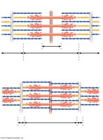
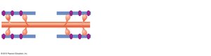
Muscle contraction occurs when calcium binds to regulatory proteins on actin, exposing myosin-binding sites. Myosin heads attach, pivot, and pull actin filaments toward the center of the sarcomere. ATP provides energy for this process.
Calcium triggers exposure of binding sites.
Myosin heads attach and pivot, causing contraction.
ATP is required for myosin head detachment and re-cocking.
Energy for Muscle Contraction
ATP Generation Pathways
Muscle fibers use three main pathways to generate ATP:
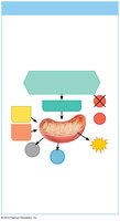
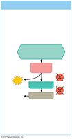
Direct phosphorylation by creatine phosphate (CP): Fastest, supplies energy for about 15 seconds.
Aerobic respiration: Occurs in mitochondria, produces about 32 ATP per glucose, requires oxygen.
Anaerobic glycolysis: Produces 2 ATP per glucose, converts pyruvic acid to lactic acid, does not require oxygen.
Muscle Fatigue and Oxygen Deficit
Causes and Recovery
Muscle fatigue occurs after prolonged activity due to ion imbalances, oxygen deficit, lactic acid accumulation, and decreased ATP supply. Recovery involves rapid, deep breathing to repay the oxygen deficit.
Muscles and Body Movements
Muscle Attachments
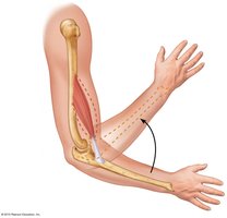
Muscles are attached to bones at two points:
Origin: Attachment to an immovable bone.
Insertion: Attachment to a movable bone.
Types of Body Movements
Flexion: Decreases joint angle, brings bones closer (e.g., bending elbow).
Extension: Increases joint angle, straightens bones (e.g., straightening knee).
Hyperextension: Extension beyond 180°.
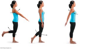
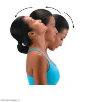
Rotation: Movement around longitudinal axis (e.g., shaking head "no").
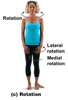
Abduction: Movement away from midline.
Adduction: Movement toward midline.
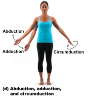
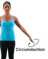
Circumduction: Circular movement combining flexion, extension, abduction, and adduction.
Special Movements
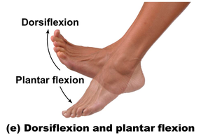
Dorsiflexion: Lifting foot toward shin.
Plantar flexion: Pointing toes downward.
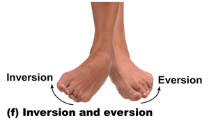
Inversion: Turning sole of foot medially.
Eversion: Turning sole of foot laterally.
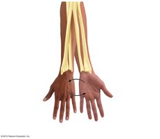
Supination: Forearm rotates laterally, palm faces anteriorly.
Pronation: Forearm rotates medially, palm faces posteriorly.
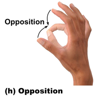
Opposition: Thumb touches tips of other fingers.