 Back
The Muscular System: Structure, Function, and Major Muscles
Study Guide - Smart Notes
 Tailored notes based on your materials, expanded with key definitions, examples, and context.
The Muscular System
Introduction to the Muscular System
The muscular system is a fundamental component of human anatomy, responsible for movement, stability, and various physiological functions. Skeletal muscles, the most prominent type, are attached to bones and facilitate voluntary movements.
Structure of Skeletal Muscle
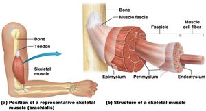
Skeletal muscle is composed of muscle cells (fibers), connective tissue, blood vessels, and nerves. The organization of these components ensures efficient contraction and communication.
Endomysium: Thin connective tissue surrounding each muscle fiber.
Perimysium: Connective tissue that bundles muscle fibers into fascicles.
Epimysium: Outer fibrous connective tissue wrapping that encloses all fascicles of a muscle.
Blood Vessels and Nerves: Supply oxygen, nutrients, and coordinate contraction.
Muscle Terminology and Naming
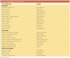
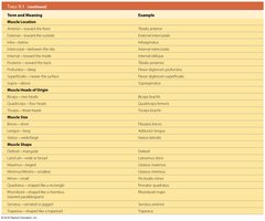
Muscles are named based on their location, action, shape, size, and orientation. Understanding these terms is essential for identifying and describing muscles.
Location: Directional terms and anatomical structures (e.g., brachialis for the arm).
Action: Flexors, extensors, adductors, abductors, levators, etc.
Shape and Size: Deltoid (triangle-shaped), maximus (largest), minimus (smallest).
Fiber Orientation: Rectus (straight), oblique (angled).
Term and Meaning | Example |
|---|---|
Abdominis – abdominal area | Rectus abdominis |
Brachii – arm area | Biceps brachii |
Flexor – muscle that flexes | Flexor digitorum longus |
Extensor – muscle that extends | Extensor digitorum |
Maximus – largest | Gluteus maximus |
Minimus – smallest | Gluteus minimus |
Deltoid – triangular shape | Deltoid |
Rectus – straight | Rectus femoris |
Oblique – angled | External oblique |
Transversus – transverse | Transversus abdominis |
Longus – long | Adductor longus |
Brevis – short | Adductor brevis |
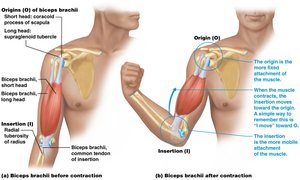
Origin – fixed attachment | Biceps brachii origin |
Insertion – movable attachment | Biceps brachii insertion |
Additional info: See images for more examples. |
Motor Units and Muscle Function
A motor unit consists of a single motor neuron and all the muscle fibers it innervates. The number of fibers in a motor unit determines the precision of movement.
Small motor units: Few muscle fibers, allow for precise movements (e.g., fingers).
Large motor units: Many muscle fibers, allow for powerful but less precise movements (e.g., thigh muscles).
Form follows function: The shape, size, and arrangement of muscle fibers contribute to the muscle's function.
Functions of Skeletal Muscles
Skeletal muscles perform various functions beyond movement, including heat generation, respiration, and control of body openings.
Movement: Muscles contract to move bones at joints.
Heat Production: Muscle contractions generate heat, aiding in temperature regulation.
Respiration: The diaphragm is essential for breathing.
Facial Expression: Muscles attached to facial skin allow for expressions.
Swallowing: Muscles in the throat assist with swallowing.
Sphincters: Control opening and closing of body passages.
Functional Groups of Muscles
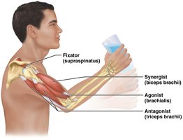
Muscles often work in groups to perform movements. Each muscle in a group has a specific role:
Agonist (Prime Mover): Provides the main force for a movement.
Antagonist: Opposes the action of the agonist, allowing for control and modulation.
Synergist: Assists the agonist by providing additional force and stabilizing joints.
Fixator: Stabilizes the origin of the agonist, increasing movement efficiency and preventing injury.
Muscle Origin and Insertion
Each skeletal muscle has a distinct origin and insertion:
Origin: The fixed, anchoring point on a bone, typically not involved in movement.
Insertion: The movable end of the muscle, attached to a bone or structure that moves during contraction.
Major Muscles of the Body
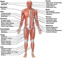
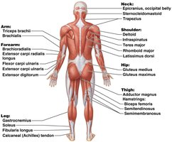
The human body contains numerous muscles, each with specific functions and anatomical locations. Muscles are grouped by region for easier study.
Head: Temporalis, Masseter
Shoulder: Deltoid
Arm: Triceps brachii, Biceps brachii, Brachialis
Forearm: Pronator teres, Flexor carpi radialis, Extensor digitorum
Thigh: Quadriceps femoris, Adductor longus, Gracilis
Leg: Gastrocnemius, Soleus, Tibialis anterior
Muscles of Facial Expression
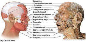
Facial muscles insert into the skin and connective tissue, allowing for subtle changes in facial structure and expression.
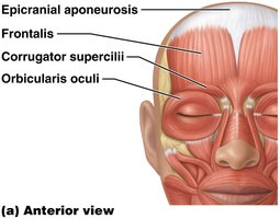
Epicranial aponeurosis: Connects frontalis and occipitalis muscles.
Orbicularis oculi: Closes the eyelids.
Levator palpebrae superioris: Raises the upper eyelid.
Corrugator supercilii: Draws eyebrows together.
Muscles involved in mouth movement include:
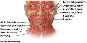
Zygomaticus major and minor: Elevate the corners of the mouth.
Levator labii superioris: Elevates the upper lip.
Risorius: Draws the mouth laterally.
Orbicularis oris: Controls movements for eating, drinking, and speaking.
Muscles of the Head and Neck
These muscles facilitate chewing, swallowing, and movement of the head and neck.
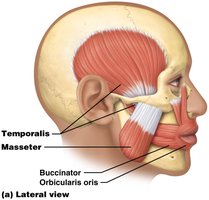
Masseter and Temporalis: Elevate the mandible for biting and chewing.
Tongue Muscles: Genioglossus, hyoglossus, styloglossus move food and aid swallowing.
Pharyngeal Muscles: Sternohyoid, sternothyroid, omohyoid, thyrohyoid, pharyngeal constrictors initiate swallowing.
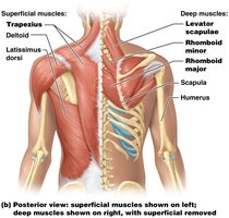
Neck Muscles: Sternocleidomastoid, scalenes, trapezius, splenius capitis, splenius cervicis, semispinalis capitis move the head and neck.
Muscles of the Trunk
Trunk muscles are grouped by proximity and function, including respiration and abdominal muscles.
Respiratory Muscles: Diaphragm, internal intercostals, external intercostals.
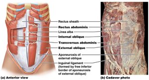
Abdominal Muscles: Rectus abdominis, external obliques, internal obliques, transversus abdominis.
Linea alba: Connective tissue separating left and right rectus muscles.
Muscles of the Shoulder and Arm
These muscles are associated with the pectoral girdle, arm, forearm, and hand, enabling a wide range of movements.
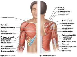
Deltoid: Abducts the arm.
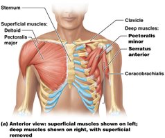
Pectoralis major: Flexes, adducts, and rotates the arm.
Serratus anterior: Protracts the scapula.
Rotator cuff group: Supraspinatus, infraspinatus, teres minor, subscapularis stabilize the shoulder.
Biceps brachii, brachialis, brachioradialis: Flex the elbow.
Triceps brachii, anconeus: Extend the elbow.
Muscles of the Arm, Forearm, and Hand
Muscles in these regions are organized by their origin, action, and insertion.
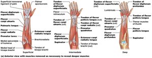
Wrist Flexors (anterior forearm): Flexor carpi radialis, palmaris longus, flexor carpi ulnaris.
Finger and Thumb Flexors (deep anterior forearm): Flexor digitorum superficialis, flexor digitorum profundus, flexor pollicis longus.
Pronators: Pronator teres, pronator quadratus.
Supinator: Supinator muscle.
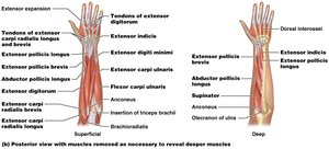
Wrist Extensors (posterior forearm): Extensor carpi radialis longus, extensor carpi radialis brevis, extensor carpi ulnaris.
Finger and Thumb Extensors: Extensor digitorum, extensor indicis, extensor digiti minimi, extensor pollicis longus, extensor pollicis brevis.
Muscles That Move the Thigh and Knee
Muscles of the pelvis and thigh are essential for locomotion and support. The pelvis is firmly attached to the axial skeleton, allowing thigh and leg muscles to focus on movement.
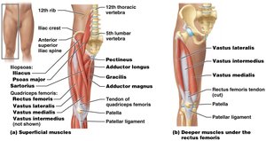
Anterior thigh muscles: Iliopsoas, sartorius (flex thigh at hip).
Quadriceps femoris group: Rectus femoris, vastus lateralis, vastus medialis, vastus intermedius (extend leg at knee).
Medial thigh muscles: Adductor magnus, adductor longus, adductor brevis, pectineus, gracilis (adduct thigh, assist with hip and knee flexion).
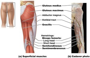
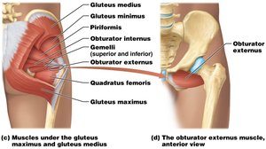
Gluteal group: Gluteus maximus, gluteus medius, gluteus minimus (extend, abduct, rotate thigh).
Hamstrings: Biceps femoris, semitendinosus, semimembranosus (flex leg at knee).
Deep posterior muscles: Piriformis, obturator internus, gemelli, quadratus femoris, obturator externus (laterally rotate thigh).
Muscles That Move the Ankle and Foot
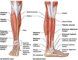
Muscles in the leg and foot are responsible for movements such as dorsiflexion, plantar flexion, inversion, eversion, and toe movements.
Tibialis anterior, extensor digitorum longus: Dorsiflex the foot at the ankle.
Flexor hallucis longus: Extends the great toe, assists with dorsiflexion.
Triceps surae group: Soleus, gastrocnemius, plantaris (plantar flex the foot).
Fibularis longus, fibularis brevis: Evert the foot.
Tibialis posterior, flexor hallucis longus, flexor digitorum longus: Invert the foot, flex toes.
Intrinsic foot muscles: Flexor digitorum brevis, lumbricals, plantar interossei, dorsal interossei (move toes).
Clinical Applications: Muscle Injuries and Disorders
Muscle injuries such as rotator cuff tears and calcaneal tendon ruptures are common in athletes and older adults. Prevention and treatment involve targeted exercises, stretching, and medical intervention when necessary.
Rotator cuff injuries: Caused by forceful overhead movements; symptoms include pain and loss of movement.
Calcaneal tendon injuries: Common in sports; treatment includes rest, physical therapy, or surgery.
Muscle knots (myofascial trigger points): Painful contractions of muscle fibers; treated with massage, medication, and stretching.
Back pain: Often due to muscle spasm, disc issues, or ligament injury; prevention includes exercise and proper lifting techniques.
Additional info: For detailed muscle charts and clinical case studies, refer to textbook appendices and clinical sections.
