 Back
The Muscular System: Structure, Function, and Major Muscles
Study Guide - Smart Notes
 Tailored notes based on your materials, expanded with key definitions, examples, and context.
The Muscular System
Overview of the Muscular System
The muscular system is a fundamental component of human anatomy, responsible for movement, posture, and various physiological functions. It consists primarily of skeletal muscles, which are under voluntary control and attach to bones via tendons.
Structure of Skeletal Muscle
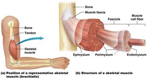
Skeletal muscle is composed of muscle cells (fibers), connective tissue, blood vessels, and nerves. The organization of these components allows for efficient contraction and force transmission.
Endomysium: Thin connective tissue surrounding each muscle fiber.
Perimysium: Connective tissue that bundles muscle fibers into fascicles.
Epimysium: Outer fibrous connective tissue wrapping that encloses all fascicles of a muscle.
Blood Vessels and Nerves: Supply oxygen, nutrients, and coordinate contraction.
Motor Unit: A motor unit consists of a single motor neuron and all the muscle fibers it innervates. Smaller motor units allow for more precise movements.
Form Follows Function: The shape, size, and arrangement of muscle fibers contribute to the specific function of each muscle.
Naming Muscles
Muscles are named based on their location, appearance, size, position, attachments, and actions. Historical names may also persist.
Location: Directional terms and word roots (e.g., brachialis for arm).
Attachment: Anatomical structures where muscles originate or insert.
Action: Flexors, extensors, adductors, abductors, levators, etc.
Common Terms in Muscle Anatomy
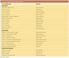
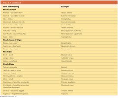
Muscle terminology is standardized to describe regions, actions, locations, origins, fiber orientation, size, and shape.
Term and Meaning | Example |
|---|---|
Abdominis – abdominal area | Rectus abdominis |
Brachii – arm | Biceps brachii |
Femoris – thigh | Rectus femoris |
Flexor – muscle that flexes | Flexor digitorum longus |
Extensor – muscle that extends | Extensor digitorum |
Longus – long | Adductor longus |
Brevis – short | Adductor brevis |
Deltoid – triangular | Deltoid |
Maximus – largest | Gluteus maximus |
Minimus – smallest | Gluteus minimus |
Oblique – angled | External oblique |
Transversus – transverse | Transversus abdominis |
Rectus – straight | Rectus abdominis |
Functions of Skeletal Muscles
Movement and Other Functions
Skeletal muscles are responsible for movement of bones at joints, but also perform other vital functions:
Heat Generation: Muscle contractions produce heat, aiding in body temperature regulation.
Respiration: The diaphragm is essential for breathing.
Facial Expression: Muscles attached to facial skin allow for expression.
Swallowing: Muscles in the throat assist with swallowing.
Sphincters: Skeletal muscle sphincters control body openings.
Functional Groups of Muscles
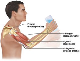
Muscles often work in groups to perform movements. The roles within these groups are defined as follows:
Agonist (Prime Mover): Provides the main force for a movement.
Antagonist: Opposes the action of the agonist, allowing for control and modulation.
Synergist: Assists the agonist by providing additional force and stabilizing joints.
Fixator: Stabilizes the origin of the agonist, increasing movement efficiency and preventing injury.
Muscle Origin and Insertion
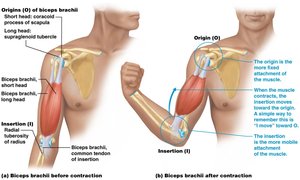
Each skeletal muscle has a distinct origin and insertion:
Origin: The anchoring point, typically on a bone, not directly involved in movement.
Insertion: The moving end, attached to a bone or structure, usually on the far side of a joint.
Major Muscles of the Body
Superficial Muscles: Anterior and Posterior Views
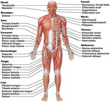
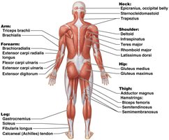
The human body contains numerous muscles, each with specific functions and anatomical locations. Understanding their arrangement is essential for clinical and academic purposes.
Muscles of Facial Expression
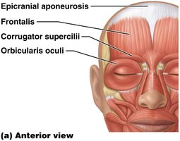
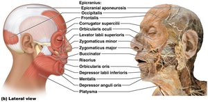
Facial muscles insert into the skin and connective tissue, allowing for subtle changes in facial structure and expression. The epicranial aponeurosis connects the frontalis and occipitalis muscles, forming the epicranius, which raises the eyebrows and forehead skin.
Orbicularis oculi: Closes the eyelids.
Levator palpebrae superioris: Elevates the upper eyelid.
Corrugator supercilii: Draws eyebrows downward and medially.
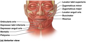
Muscles involved in mouth movement include:
Zygomaticus major and minor: Elevate the corners of the mouth.
Levator labii superioris: Elevates the upper lip.
Risorius: Draws the corner of the mouth laterally.
Orbicularis oris: Controls movements for eating, drinking, and speaking.
Muscles of the Head and Neck
Head and neck muscles facilitate chewing, swallowing, and movement of the head and neck.
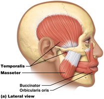
Masseter and Temporalis: Elevate the mandible for biting and chewing.
Tongue Muscles: Genioglossus, hyoglossus, and styloglossus move food during chewing and swallowing.
Pharyngeal Muscles: Sternohyoid, sternothyroid, omohyoid, thyrohyoid, and pharyngeal constrictors initiate swallowing.
Neck Muscles: Sternocleidomastoid, scalenes, trapezius, splenius capitis, splenius cervicis, and semispinalis capitis move the head and neck.
Muscles of the Trunk
Abdominal Muscles
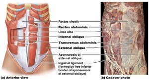
The abdominal muscles are grouped by anatomical proximity and function. They include:
Rectus abdominis: Enclosed in the rectus sheath, separated by the linea alba.
External and Internal Obliques: Contribute to trunk rotation and lateral flexion.
Transversus abdominis: Compresses abdominal contents.
Muscles of the Shoulder and Arm
Muscles Associated with the Pectoral Girdle, Arm, Forearm, and Hand
These muscles are responsible for movements of the shoulder, arm, forearm, and hand.
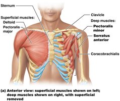
Deltoid: Abducts the arm.
Pectoralis major: Flexes, adducts, and medially rotates the arm.
Serratus anterior: Protracts the scapula.
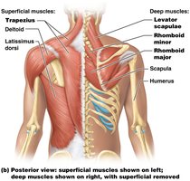
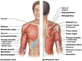
Muscles that move the humerus include pectoralis major, latissimus dorsi, teres major, coracobrachialis, teres minor, supraspinatus, infraspinatus, and subscapularis (rotator cuff group).
Muscles that move the forearm at the elbow:
Biceps brachii, brachialis, brachioradialis: Flex the elbow.
Triceps brachii, anconeus: Extend the elbow.
Muscles of the Arm, Forearm, and Hand
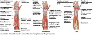
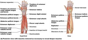
Muscles are arranged by origin, action, and insertion. Wrist flexors and extensors, finger and thumb flexors and extensors, and muscles involved in pronation and supination are key groups.
Wrist Flexors: Flexor carpi radialis, palmaris longus, flexor carpi ulnaris.
Finger Flexors: Flexor digitorum superficialis, flexor digitorum profundus, flexor pollicis longus.
Pronators: Pronator teres, pronator quadratus.
Supinator: Supinator muscle.
Wrist Extensors: Extensor carpi radialis longus, extensor carpi radialis brevis, extensor carpi ulnaris.
Finger Extensors: Extensor digitorum, extensor indicis, extensor digiti minimi, extensor pollicis longus, extensor pollicis brevis.
Muscles That Move the Thigh and Knee
Anterior, Medial, and Posterior Muscles
The pelvis is firmly affixed to the axial skeleton, allowing nearly all muscles in the pelvis and thigh to move the thigh and leg.
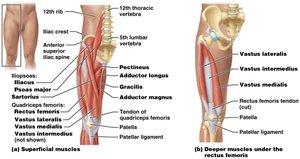
Anterior Muscles: Iliopsoas and sartorius flex the thigh at the hip. Quadriceps femoris group (rectus femoris, vastus lateralis, vastus medialis, vastus intermedius) extends the leg at the knee.
Medial Muscles: Adductor magnus, adductor longus, adductor brevis, pectineus, and gracilis adduct the thigh and assist with hip and knee flexion.
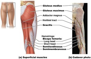
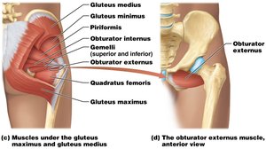
Posterior Muscles: Gluteal group (gluteus maximus, medius, minimus) extends, abducts, and rotates the thigh. Hamstrings (biceps femoris, semitendinosus, semimembranosus) flex the leg at the knee. Deep posterior muscles (piriformis, obturator internus, gemelli, quadratus femoris, obturator externus) laterally rotate the thigh.
Muscles That Move the Ankle and Foot
Muscles of the Leg and Foot
Muscles that move the foot and toes are grouped by their location and action:
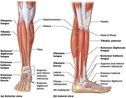
Anterior Leg: Tibialis anterior and extensor digitorum longus dorsiflex the foot at the ankle.
Flexor hallucis longus: Assists with dorsiflexion and extends the great toe.
Triceps surae group: Soleus, gastrocnemius, and plantaris plantar flex the foot at the ankle.
Lateral Leg: Fibularis longus and brevis allow for foot eversion.
Deep Muscles: Tibialis posterior and anterior allow for foot inversion; flexor hallucis longus flexes the great toe; flexor digitorum longus flexes toes 2–5.
Intrinsic Foot Muscles: Flexor digitorum brevis, lumbricals, plantar interossei, and dorsal interossei move the toes.
Clinical Applications
Muscle Knots (Myofascial Trigger Points)
Muscle knots are groups of muscle fibers or fascicles that contract painfully and will not relax. They can cause severe pain and stress on joints and connective tissue. Causes include microscopic tears from repetitive exercise, sudden muscle use, poor posture, sedentary lifestyle, diet, and ion imbalance.
Palpation: Used to identify trigger points.
Treatment: Massage therapy, anti-inflammatory medications, muscle relaxants, and regular stretching.
Prevention: Moderate exercise and a healthy diet.
Back Pain
Back pain is a common medical problem, often due to degenerated or herniated intervertebral discs, muscle spasm, or ligament injury. Risk factors include age, obesity, poor fitness, genetics, and occupation. Prevention involves healthy practices; treatment ranges from self-care to medical intervention.
Rotator Cuff Injuries
The rotator cuff group stabilizes the shoulder joint during forceful movements. Injuries range from inflammation to acute tears requiring surgery. Prevention involves targeted exercises; physical therapy is important after injury.
Calcaneal (Achilles) Tendon Injuries
Calcaneal tendon injuries are common in sports and activities involving running and directional changes. The tendon is vulnerable due to the lever mechanics of the foot. Treatment may be nonsurgical or surgical; prevention includes stretching and gradual increase in activity intensity.
Kegel Exercises
Pelvic floor exercises strengthen pelvic floor muscles, reinforcing sphincters and preventing pelvic organ prolapse. Risk factors include childbirth, age, weight gain, hormonal changes, and changes in reproductive structures.
Additional info: The notes above expand on brief points with academic context, definitions, and examples to ensure completeness and clarity for college-level Anatomy & Physiology students.
