 Back
BackThe Muscular System: Structure, Function, and Physiology
Study Guide - Smart Notes
 Tailored notes based on your materials, expanded with key definitions, examples, and context.
The Muscular System
Functions of Muscle
The muscular system is essential for movement, stability, and regulation of bodily functions. Muscles generate force and produce motion, maintain posture, regulate openings, and contribute to thermoregulation.
Movement: Muscles contract to move bones and body parts.
Opening and Closing: Sphincter muscles control passageways (e.g., digestive tract).
Stabilizing and Containing: Muscles stabilize joints and contain organs.
Temperature Control: Muscle activity generates heat, accounting for up to 85% of body heat.
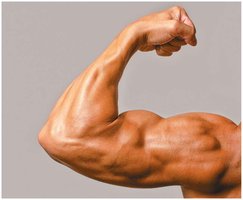
Characteristics of Muscle Tissue
Muscle tissue possesses unique properties that enable its function:
Excitability: Ability to respond to stimuli and generate electrical impulses.
Contractility: Ability to shorten forcibly when stimulated.
Extensibility: Ability to stretch beyond resting length.
Elasticity: Ability to return to original shape after stretching or contracting.
Types of Muscle Tissue
Skeletal, Cardiac, and Smooth Muscle
There are three main types of muscle tissue, each with distinct structure and function:
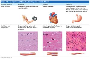
Skeletal Muscle: Voluntary, striated, multinucleated cells attached to bones for movement.
Cardiac Muscle: Involuntary, striated, branched cells found in the heart, responsible for pumping blood.
Smooth Muscle: Involuntary, non-striated, spindle-shaped cells found in walls of hollow organs.
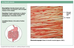
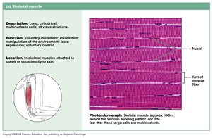
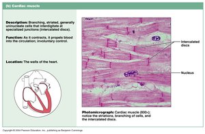
Comparison Table: Skeletal, Cardiac, and Smooth Muscle
Characteristic | Skeletal | Cardiac | Smooth |
|---|---|---|---|
Body Location | Attached to bones or skin | Walls of the heart | Walls of hollow organs |
Cell Shape & Appearance | Long, cylindrical, multinucleated, striated | Branched, striated, single or two nuclei | Spindle-shaped, non-striated, single nucleus |
Control | Voluntary | Involuntary | Involuntary |
Organization of Skeletal Muscle
Structural Hierarchy
Skeletal muscle is organized into a hierarchy of structures, from smallest to largest:
Myofilaments: Actin and myosin proteins forming the cytoskeleton.
Sarcomere: Functional unit of contraction, composed of bands and zones.
Myofibril: Bundles of sarcomeres within a muscle cell.
Myofiber: Individual muscle cell (myocyte).
Muscle Fascicle: Bundle of muscle fibers.
Named Muscle: Entire muscle organ.
Muscle Group: Group of muscles with related function.
Connective Tissue Coverings
Connective tissue layers provide support and organization:
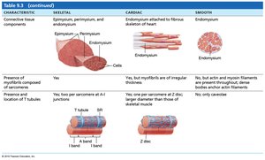
Endomysium: Surrounds each muscle fiber.
Perimysium: Surrounds each fascicle.
Epimysium: Surrounds the entire muscle.
Deep Fascia: Surrounds muscle groups and attaches to bone or tendons.
Muscle Cell (Myofiber) vs. General Cell
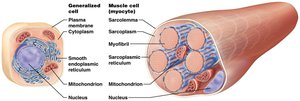
Muscle cells are specialized for contraction and differ structurally from typical cells:
Sarcoplasm: Specialized cytoplasm.
Sarcolemma: Plasma membrane of muscle cell.
Sarcoplasmic Reticulum (SR): Modified smooth ER, stores calcium ions.
Myofilaments: Actin and myosin proteins for contraction.
Microscopic Anatomy: Sarcomere and Myofilaments
The sarcomere is the fundamental contractile unit of muscle, composed of organized myofilaments:
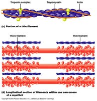
Thin Filaments (Actin): Includes G-actin (with myosin-binding sites), tropomyosin (blocks binding sites), and troponin (binds calcium).
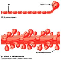
Thick Filaments (Myosin): Composed of myosin molecules with heads and tails.
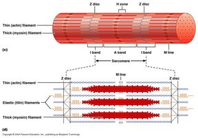
Elastic Filaments (Titin): Connects myosin to Z-disc, provides elasticity.
Sarcomere Structure and Bands
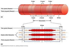
Sarcomeres are defined by Z-discs and contain distinct bands:
I Band: Contains only thin filaments.
H Zone: Contains only thick filaments.
A Band: Overlap of thick and thin filaments.
Z Disc: Boundary of sarcomere.
M Line: Center of thick filaments.
Sliding Filament Theory and Muscle Contraction
Muscle contraction occurs through the sliding filament mechanism:
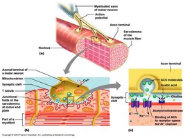
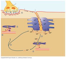
Motor neuron fires, sending an electrical impulse to the muscle cell.
Impulse travels along sarcolemma and T-tubules, triggering SR to release Ca2+.
Ca2+ binds to troponin, moving tropomyosin and exposing myosin-binding sites on actin.
Myosin heads bind to actin, pull, and cause contraction.
ATP is required for myosin head detachment and re-cocking.
Muscle Relaxation
Relaxation occurs when:
Acetylcholinesterase breaks down acetylcholine in the synaptic cleft.
ATP pumps Ca2+ back into the SR.
ATP breaks actin-myosin crossbridges.
Muscle returns to resting state.
Motor Units and Neuromuscular Junction
Motor Units
A motor unit consists of a motor neuron and all the muscle fibers it innervates. The number and size of motor units affect muscle precision and strength.
Fine Movements: Many small motor units (e.g., fingers, eyes).
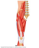
Strength Movements: Fewer, larger motor units (e.g., legs, back).
Neuromuscular Junction
The neuromuscular junction is the site where a motor neuron communicates with a muscle fiber:
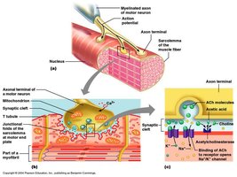
Axon Terminal: Contains synaptic vesicles with acetylcholine (ACh).
Motor End Plate: Region of sarcolemma with ACh receptors.
Synaptic Cleft: Space between neuron and muscle cell.
Muscle Tension and Contraction Types
Muscle Tension
Muscle tension is the force generated by muscle contraction. It depends on:
Frequency of stimulation
Number of fibers contracting
Length of sarcomeres
Types of Contractions
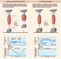
Isometric Contraction: Muscle length remains constant, tension changes (posture maintenance).
Isotonic Contraction: Muscle length changes, tension remains constant (movement).
Concentric: Muscle shortens (lifting weight).
Eccentric: Muscle lengthens (lowering weight).
Muscle Twitch, Treppe, and Tetanus
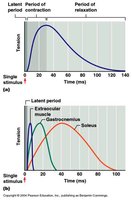
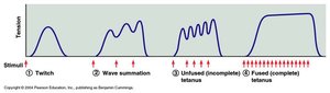
A muscle twitch is a single contraction in response to a stimulus. Treppe is a staircase effect of increasing contractions. Tetanus is sustained contraction due to rapid stimuli.
Latent Period: Ca2+ released from SR.
Contraction Period: Filaments slide.
Relaxation Period: Ca2+ pumped back into SR.
Refractory Period: Muscle cannot respond to another stimulus.
Muscle Metabolism and Fiber Types
Energy Needs and Sources
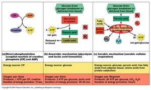
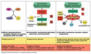
Muscles require ATP for contraction, crossbridge cycling, and ion transport. ATP is generated by:
Creatine Phosphate: Rapid, short-term energy source.
Anaerobic Glycolysis: Produces ATP and lactic acid without oxygen.
Aerobic Respiration: Produces large amounts of ATP with oxygen.
Muscle Fiber Types
Slow Oxidative Fibers (Type I): Small, fatigue-resistant, aerobic, postural muscles.
Fast Oxidative-Glycolytic Fibers (Type IIa): Intermediate, aerobic and anaerobic, moderately fatigue-resistant.
Fast Glycolytic Fibers (Type IIb): Large, powerful, anaerobic, fatigue quickly.
Levers and Mechanical Advantage in the Body
Lever Systems
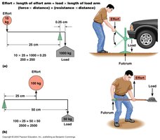
Muscles act on bones as levers, with joints as fulcrums. Levers are classified as:
First Class: Fulcrum in the middle (e.g., neck joint).
Second Class: Load in the middle (e.g., foot arch).
Third Class: Effort in the middle (e.g., elbow joint).
Smooth and Cardiac Muscle
Smooth Muscle
Smooth muscle lines hollow organs and is responsible for involuntary movements such as peristalsis and sphincter control. Cells are spindle-shaped, non-striated, and have a single nucleus.
Contraction: Initiated by Ca2+ binding to calmodulin, activating myosin light chain kinase.
Subtypes: Single-unit (gap junctions, contracts as a unit) and multi-unit (independent cells, precision control).
Cardiac Muscle
Cardiac muscle is found only in the heart. Cells are branched, striated, and connected by intercalated discs (gap junctions and desmosomes). Contractions are involuntary and autorhythmic.
Comparison Table: Skeletal, Cardiac, and Smooth Muscle (Summary)
Feature | Skeletal | Cardiac | Smooth |
|---|---|---|---|
Striations | Yes | Yes | No |
Control | Voluntary | Involuntary | Involuntary |
Location | Bones | Heart | Hollow organs |
Cell Shape | Long, cylindrical | Branched | Spindle-shaped |
Additional info: These notes expand on the original content by providing definitions, examples, and structured tables for comparison. All images included are directly relevant to the adjacent explanations and reinforce key concepts in muscle anatomy and physiology.
