 Back
BackThe Muscular System: Structure, Function, and Major Muscles
Study Guide - Smart Notes
 Tailored notes based on your materials, expanded with key definitions, examples, and context.
The Muscular System
Overview of Muscle Tissue
The muscular system is composed of all contractile tissues in the body, including skeletal, cardiac, and smooth muscle. This chapter focuses on skeletal muscle, which is responsible for voluntary movements and is attached to bones by tendons.
Skeletal muscle: Voluntary, striated muscle attached to bones.
Cardiac muscle: Involuntary, striated muscle found in the heart.
Smooth muscle: Involuntary, non-striated muscle found in walls of hollow organs.
Key topics include how muscles interact to produce movement, criteria for naming muscles, and principles of leverage.
Interactions of Skeletal Muscles
Muscle Functional Groups
Skeletal muscles work together in groups to produce coordinated movements. Muscles can only pull, not push; thus, movements are produced by the contraction of one muscle and reversed by another.
Prime mover (agonist): Main muscle responsible for a specific movement.
Antagonist: Muscle that opposes or reverses a movement; located on the opposite side of the joint from the agonist.
Synergist: Assists the prime mover by adding force or reducing unnecessary movement.
Fixator: A type of synergist that stabilizes the origin of the prime mover.
Example: The biceps brachii is the prime mover for elbow flexion, while the triceps brachii is the antagonist.
Muscle Action and Joint Position
The action of a muscle can often be inferred by its position relative to the joint it crosses:
Muscle crossing the anterior side of a joint: produces flexion (except at the knee and ankle).
Muscle crossing the posterior side: produces extension.
Muscle crossing the lateral side: produces abduction.
Muscle crossing the medial side: produces adduction.
These generalities do not apply to the knee and ankle due to limb rotation during development.
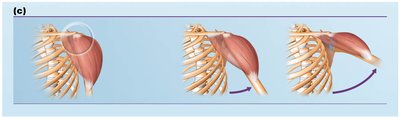
Naming Skeletal Muscles
Criteria for Naming Muscles
Muscle names often describe one or more of the following characteristics:
Location: Bone or region associated (e.g., temporalis muscle over the temporal bone).
Shape: Deltoid (triangle-shaped), trapezius (trapezoid-shaped).
Size: Maximus (largest), minimus (smallest), longus (long).
Direction of fibers: Rectus (straight), transversus (right angle), oblique (at an angle).
Number of origins: Biceps (2 origins), triceps (3 origins).
Location of attachments: Named for origin and insertion (origin first).
Action: Flexor, extensor, adductor, etc.
Several criteria can be combined, such as extensor carpi radialis longus (a long muscle that extends the wrist and is near the radius).
Fascicle Arrangements in Skeletal Muscle
Patterns of Fascicle Arrangement
Fascicles are bundles of muscle fibers, and their arrangement determines muscle shape and function. The main patterns are:
Circular: Fascicles arranged in concentric rings (e.g., orbicularis oris).
Convergent: Broad origin, fascicles converge toward a single tendon (e.g., pectoralis major).
Parallel: Fascicles parallel to the long axis (e.g., sartorius).
Fusiform: Spindle-shaped with parallel fibers (e.g., biceps brachii).
Pennate: Short fascicles attach obliquely to a central tendon. Types include unipennate, bipennate, and multipennate.
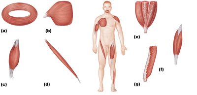
Functional Implications of Fascicle Arrangement
Range of motion: Determined by the arrangement and length of fascicles. Long, parallel fibers allow greater movement but less power.
Power: Muscles with more fibers (e.g., bipennate, multipennate) are more powerful but have less range of motion.
Major Skeletal Muscles of the Body
Muscle Groups and Identification
There are over 600 skeletal muscles in the human body, grouped by function and location. Key information for each muscle includes shape, location, origin and insertion, actions, and innervation.
Muscle actions are typically described as the insertion moving toward the origin.
Innervation refers to the major nerve supplying the muscle.
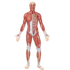
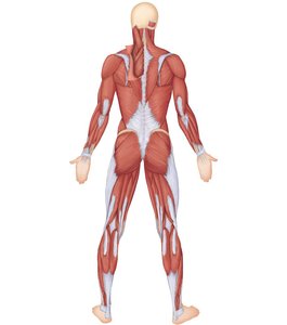
Muscles of the Head: Facial Expression
Overview
Muscles of facial expression are unique because they insert into the skin rather than bone, allowing for a wide range of nonverbal communication. All are innervated by the facial nerve (cranial nerve VII).
Divided into muscles of the scalp and face.
Examples: orbicularis oculi (closes eye), zygomaticus major (smiling), platysma (tenses neck skin).
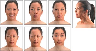
Table: Muscles of Facial Expression
Muscle | Description | Origin/Insertion | Action | Nerve Supply |
|---|---|---|---|---|
Orbicularis oculi | Thin, flat sphincter muscle of eyelids | Frontal and maxillary bones/skin of eyelid | Closes eye | Facial nerve |
Zygomaticus major | Extends from cheekbone to mouth corner | Zygomatic bone/skin at mouth angle | Raises lateral corners of mouth (smile) | Facial nerve |
Platysma | Superficial neck muscle | Fascia of chest/lower margin of mandible | Tenses skin of neck | Facial nerve |
Muscles of Mastication and Tongue Movement
Muscles of Mastication
Four pairs of muscles are responsible for chewing, all innervated by the trigeminal nerve (cranial nerve V):
Temporalis and masseter: Prime movers of jaw closure.
Pterygoids: Grinding movements.
Buccinator: Assists in chewing by compressing the cheek.
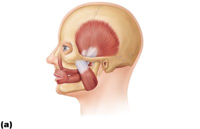
Muscles Promoting Tongue Movements
Genioglossus: Protracts tongue.
Hyoglossus: Depresses tongue.
Styloglossus: Retracts and elevates tongue.
Muscles of the Neck and Vertebral Column
Head Movements and Trunk Extension
Muscles in this region are divided into anterolateral neck muscles (move the head) and intrinsic back muscles (extend the trunk and maintain posture).
Sternocleidomastoid: Flexes and laterally rotates the head.
Scalenes: Elevate first two ribs, aid in neck flexion.
Splenius: Extend or hyperextend the head.
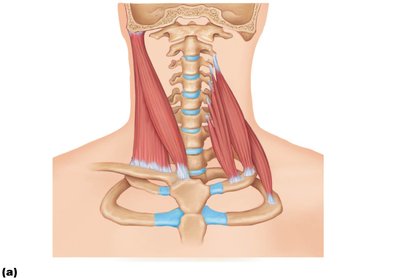
Deep Muscles of the Thorax: Breathing
Muscles of Respiration
Breathing involves two phases: inspiration (inhaling) and expiration (exhaling). The main muscles are:
Diaphragm: Most important muscle for inspiration; innervated by phrenic nerves.
External intercostals: Elevate ribs for inspiration.
Internal intercostals: Aid forced expiration.

Muscles of the Abdominal Wall
Structure and Function
The abdominal wall is formed by four paired muscles and their aponeuroses:
Rectus abdominis
External oblique
Internal oblique
Transversus abdominis
These muscles provide strength, allow lateral flexion and rotation of the trunk, and assist in functions such as urination, defecation, and childbirth.
Muscles of the Pelvic Floor and Perineum
Pelvic Diaphragm
Composed of the levator ani and coccygeus, the pelvic diaphragm supports pelvic organs, resists increased intra-abdominal pressure, and aids in fecal release.
Urogenital Diaphragm
Located anteriorly, it contains the external urethral sphincter for voluntary urination control.
Superficial Muscles of the Thorax: Movements of the Scapula and Arm
Anterior Thorax Muscles
Pectoralis minor
Serratus anterior
Subclavius
Posterior Thorax Muscles
Trapezius
Levator scapulae
Rhomboids
Muscles Crossing the Shoulder Joint: Movements of the Arm
Major Muscles
Pectoralis major
Latissimus dorsi
Deltoid
The rotator cuff muscles (supraspinatus, infraspinatus, teres minor, subscapularis) act as synergists and fixators, reinforcing the shoulder joint.
Muscles Crossing the Hip and Knee Joints
Movements of the Thigh and Leg
Hamstrings: Prime movers of thigh extension and knee flexion (biceps femoris, semitendinosus, semimembranosus).
Quadriceps femoris: Sole extensor of the knee.
Muscles of the Leg: Movements
Movements Produced
Ankle: Dorsiflexion and plantar flexion.
Intertarsal joints: Inversion and eversion of the foot.
Toes: Flexion and extension.
Additional info: For more detailed muscle tables, refer to the included tables in the original material, which provide origin, insertion, action, and innervation for each muscle group.
