 Back
BackThe Muscular System: Structure, Function, and Physiology
Study Guide - Smart Notes
 Tailored notes based on your materials, expanded with key definitions, examples, and context.
The Muscular System
Overview of Muscle Tissue
The muscular system is one of the major systems of the human body, responsible for movement, posture, and heat production. Muscle tissue is specialized for contraction and is classified into three types: skeletal, cardiac, and smooth muscle.
Skeletal muscle: Voluntary, striated muscle attached to bones for movement.
Cardiac muscle: Involuntary, striated muscle found only in the heart.
Smooth muscle: Involuntary, non-striated muscle found in walls of organs.
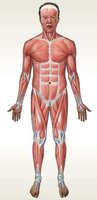
Skeletal Muscle Structure and Organization
Components of Skeletal Muscle
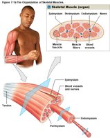
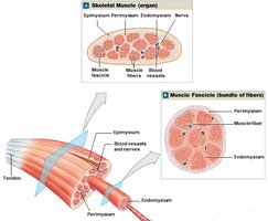
Skeletal muscles are complex organs composed of muscle tissue, connective tissues, blood vessels, and nerves. Each muscle fiber (cell) is elongated and multinucleate.
Muscle fiber: The basic cellular unit of muscle tissue.
Connective tissue layers:
Epimysium: Surrounds the entire muscle.
Perimysium: Divides muscle into fascicles (bundles of fibers).
Endomysium: Surrounds individual muscle fibers.
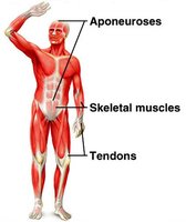
Tendon: Bundle of collagen fibers attaching muscle to bone.
Aponeurosis: Broad sheet of connective tissue connecting muscles to each other.
Microscopic Structure of Skeletal Muscle
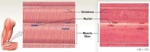
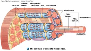
Skeletal muscle fibers are striated due to the arrangement of myofibrils, which are composed of repeating units called sarcomeres. The sarcolemma is the plasma membrane, and the sarcoplasm is the cytoplasm of the muscle fiber.
Myofibrils: Cylindrical structures containing myofilaments (actin and myosin).
Sarcomere: The functional unit of contraction, defined by Z lines.
Sarcoplasmic reticulum (SR): Specialized endoplasmic reticulum storing calcium ions.
Transverse (T) tubules: Invaginations of the sarcolemma that transmit action potentials.
Sarcomere Structure
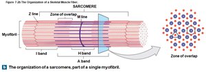
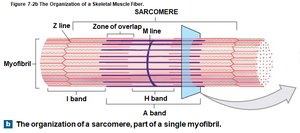
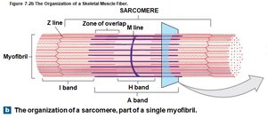
The sarcomere contains thick (myosin) and thin (actin) filaments arranged in a precise pattern, producing the striated appearance of skeletal muscle.
Z line: Boundary of each sarcomere.
M line: Center of the sarcomere, holding thick filaments together.
A band: Dark region containing thick filaments.
I band: Light region containing only thin filaments.
H band: Central region with only thick filaments (relaxed state).
Myofilaments: Thin and Thick Filaments
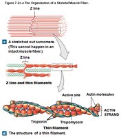
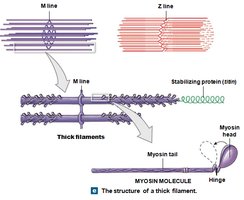
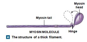
Thin filaments are primarily composed of actin, with regulatory proteins tropomyosin and troponin. Thick filaments are composed of myosin molecules with heads that interact with actin during contraction.
Actin: Contains active sites for myosin binding.
Tropomyosin: Covers active sites on actin at rest.
Troponin: Binds calcium and moves tropomyosin to expose active sites.
Myosin: Has a tail and globular head; heads form cross-bridges with actin.
Muscle Contraction
Sliding Filament Theory
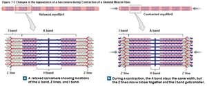
Muscle contraction occurs when thin filaments slide past thick filaments, shortening the sarcomere without changing the length of the filaments themselves.
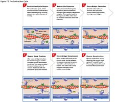
Myosin heads bind to actin, forming cross-bridges.
Heads pivot, pulling thin filaments toward the center of the sarcomere.
ATP is required for cross-bridge detachment and re-cocking of the myosin head.
Neuromuscular Junction and Excitation-Contraction Coupling
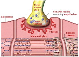
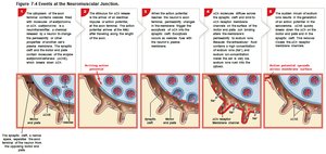
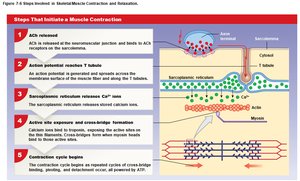
The neuromuscular junction (NMJ) is the site where a motor neuron communicates with a skeletal muscle fiber. Acetylcholine (ACh) is released, triggering an action potential in the muscle fiber, leading to calcium release from the SR and muscle contraction.
Motor end plate: Region of sarcolemma with ACh receptors.
Synaptic cleft: Gap between neuron and muscle fiber.
Excitation-contraction coupling: Sequence linking action potential to contraction.
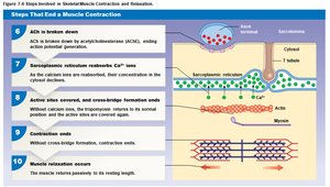
The Contraction Cycle
The contraction cycle involves repeated formation and breaking of cross-bridges between actin and myosin, powered by ATP hydrolysis.
Muscle Tension and Control
Muscle Twitch and Tension Development
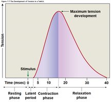
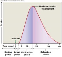
A muscle twitch is a single contraction-relaxation cycle in a muscle fiber. The amount of tension produced depends on the number of cross-bridges formed and the frequency of stimulation.
Latent period: Time between stimulus and contraction onset.
Contraction phase: Tension rises to peak.
Relaxation phase: Tension falls to resting level.
Summation and Tetanus
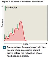
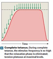
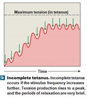
Summation occurs when stimuli arrive before the muscle has completely relaxed, increasing tension. Tetanus is a sustained contraction resulting from high-frequency stimulation.
Incomplete tetanus: Partial relaxation between stimuli.
Complete tetanus: No relaxation; maximum tension achieved.
Motor Units and Recruitment
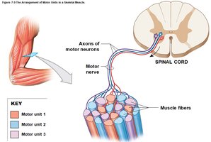
A motor unit consists of a single motor neuron and all the muscle fibers it controls. Recruitment is the process of increasing the number of active motor units to produce greater tension.
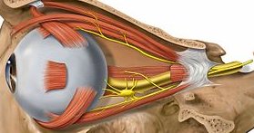
Small motor units: Fine control (e.g., eye muscles).
Large motor units: Gross movements (e.g., leg muscles).
Muscle Tone and Atrophy
Muscle tone is the resting tension in skeletal muscle, important for posture and joint stability. Atrophy is the loss of muscle mass due to lack of stimulation.
Isotonic contraction: Muscle changes length (e.g., lifting a book).
Isometric contraction: Muscle length remains the same (e.g., pushing against a wall).
Muscle Metabolism and Fatigue
ATP and Energy Storage
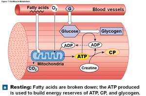
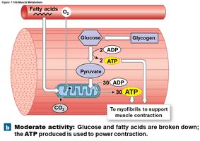
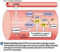
Muscle contraction requires ATP, which is generated by aerobic metabolism, anaerobic glycolysis, and stored as creatine phosphate (CP).
Aerobic metabolism: Requires oxygen, produces most ATP at rest.
Anaerobic glycolysis: Occurs without oxygen, produces less ATP but is faster.
Creatine phosphate: Stores energy to rapidly regenerate ATP.
Muscle Fatigue and Recovery
Muscle fatigue occurs when a muscle can no longer contract efficiently, often due to depletion of energy reserves or accumulation of metabolic byproducts. Recovery involves restoring energy reserves, removing lactic acid, and repaying oxygen debt.
Types of Muscle Fibers
Fast-Twitch vs. Slow-Twitch Fibers
Fast-twitch fibers: Large, powerful, fatigue quickly, rely on glycolysis.
Slow-twitch fibers: Smaller, contract slowly, fatigue-resistant, rely on aerobic metabolism and myoglobin for oxygen storage.
Muscles vary in the proportion of fiber types, affecting their color and function.
Cardiac and Smooth Muscle Tissue
Cardiac Muscle
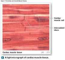
Cardiac muscle is found only in the heart, is striated, and contracts involuntarily. Cells are connected by intercalated discs, allowing coordinated contraction.
Smooth Muscle
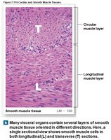
Smooth muscle is found in the walls of hollow organs, is non-striated, and contracts involuntarily. It can contract over a wide range of lengths and is regulated by neural, hormonal, or automatic mechanisms.
Comparison of Muscle Tissue Types
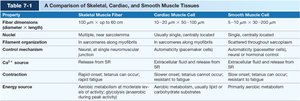
Property | Skeletal Muscle | Cardiac Muscle | Smooth Muscle |
|---|---|---|---|
Striations | Yes | Yes | No |
Control | Voluntary | Involuntary | Involuntary |
Location | Attached to bones | Heart | Walls of organs |
Cell shape | Long, cylindrical | Branched | Spindle-shaped |
Nuclei | Multiple, peripheral | Single, central | Single, central |
Muscle Actions and Naming
Origin, Insertion, and Action
Muscles are described by their origin (stationary attachment), insertion (moving attachment), and action (movement produced). Actions are classified as flexion, extension, abduction, adduction, etc.
Muscle Roles
Prime mover (agonist): Main muscle responsible for movement.
Antagonist: Opposes the prime mover.
Synergist: Assists the prime mover.
Fixator: Stabilizes the origin of the prime mover.
Muscle Naming
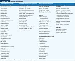
Muscles are named based on location, shape, size, direction of fibers, number of origins, and action.
Major Skeletal Muscles
Axial Muscles
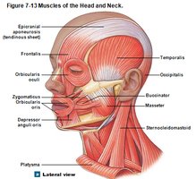
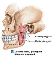
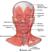
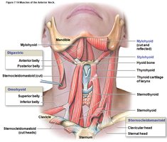
Muscles of the head and neck: Control facial expressions, mastication, and head movement.
Muscles of the spine: Maintain posture and move the vertebral column.
Muscles of the trunk: Involved in breathing, supporting abdominal organs, and moving the trunk.
Muscles of the pelvic floor: Support pelvic organs and control openings.
Appendicular Muscles
Muscles of the shoulder and upper limbs: Move the pectoral girdle, arm, forearm, wrist, hand, and fingers.
Muscles of the pelvic girdle and lower limbs: Move the thigh, leg, foot, and toes.
Effects of Aging and System Integration
Aging Effects
Muscle fibers decrease in size and number.
Muscles become less elastic and more fibrous.
Exercise tolerance and thermoregulation decrease.
Recovery from injury is slower.
System Integration
The muscular system works closely with the skeletal, cardiovascular, respiratory, integumentary, nervous, and endocrine systems to produce movement, maintain posture, and regulate body temperature.
