Back
BackThe Muscular System: Structure, Function, and Organization
Study Guide - Smart Notes
 Tailored notes based on your materials, expanded with key definitions, examples, and context.
The Muscular System
Overview of Skeletal Muscles
The muscular system is essential for movement, posture, and various physiological functions. Skeletal muscles are composed of muscle cells, connective tissues, blood vessels, and nerves, all working together to produce voluntary movements.
Skeletal Muscle Structure
Connective Tissue Organization
Skeletal muscle is not made of muscle cells alone; it also contains connective tissue, blood vessels, and nerves. The connective tissue layers organize and support muscle fibers, transmit force, and connect muscles to bones via tendons.
Endomysium: Surrounds each individual muscle cell (fiber).
Perimysium: Encloses bundles of muscle cells called fascicles.
Epimysium: Encloses all fascicles to form the complete muscle.
Fascia: Outermost layer, continuous with the epimysium, supports the muscle and attaches it to bone.
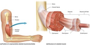
Additional info: Connective tissue layers merge to form tendons, ensuring the muscle pulls as a unit.
Motor Units
Definition and Function
A motor unit consists of a single motor neuron and all the muscle fibers it innervates. The number of muscle fibers per motor unit determines the precision of movement.
Fewer muscle fibers per motor unit = more precise movements (e.g., finger muscles).
More muscle fibers per motor unit = less precise, more powerful movements (e.g., thigh muscles).
Fascicle Arrangement and Muscle Shapes
Types of Fascicle Arrangements
The arrangement of fascicles (bundles of muscle fibers) affects both the appearance and function of skeletal muscles. "Form follows function" is a key principle in muscle anatomy.
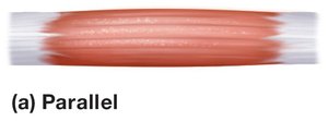
Parallel: Fascicles run parallel to the long axis of the muscle. Example: Sartorius.
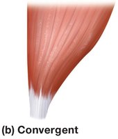
Convergent: Broad origin, fascicles converge to a single tendon. Example: Pectoralis major.
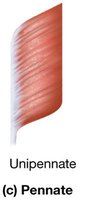
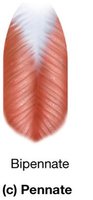
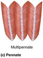
Pennate: Fascicles attach at an angle to a central tendon. Variations include unipennate, bipennate, and multipennate. Examples: Flexor pollicis longus (unipennate), rectus femoris (bipennate), deltoid (multipennate).
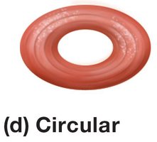
Circular (Sphincter): Fascicles arranged in concentric rings. Example: Orbicularis oculi.
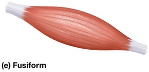
Fusiform: Muscle belly is thicker than the tapered ends. Example: Biceps brachii.
Naming Skeletal Muscles
Muscle Naming Conventions
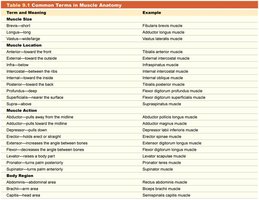
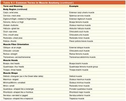
Skeletal muscles are named based on their appearance, size, location, attachments, and actions. Many names use Latin or Greek roots and may reference the muscle's function or anatomical position.
Location: e.g., tibialis anterior (front of tibia)
Size: e.g., gluteus maximus (largest gluteal muscle)
Shape: e.g., deltoid (triangular shape)
Direction of fibers: e.g., rectus abdominis (straight fibers)
Number of origins: e.g., biceps brachii (two origins)
Action: e.g., flexor carpi radialis (flexes wrist)
Term and Meaning | Example |
|---|---|
Muscle Size | Gluteus maximus |
Muscle Location | Tibialis anterior |
Muscle Shape | Deltoid |
Muscle Action | Flexor carpi radialis |
Number of Origins | Biceps brachii |
Fiber Orientation | Rectus abdominis |
Functions of Skeletal Muscles
Main and Accessory Functions
The primary function of skeletal muscle is to produce movement (action). Other important functions include:
Generating heat (thermogenesis)
Respiratory movements (e.g., diaphragm contraction)
Facial expression
Swallowing
Controlling body openings (sphincters)
Functional Groups of Muscles
Muscle Roles in Movement
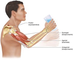
Muscles work in groups to produce coordinated movements. The main roles include:
Agonist (Prime Mover): Main muscle responsible for a movement.
Antagonist: Opposes the action of the agonist.
Synergist: Assists the agonist by adding force or reducing unwanted movement.
Fixator: Stabilizes the origin of the agonist for efficient movement.
Muscle Attachments: Origin and Insertion
Definitions and Importance
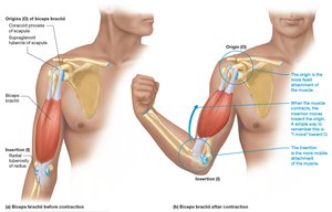
Skeletal muscles attach to bones at two main points:
Origin: The fixed, less movable attachment point.
Insertion: The movable attachment point, where movement occurs during contraction.
Lever Systems in the Body
Classes of Levers
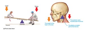
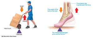
Muscles and bones act together as levers to produce movement. There are three classes of levers, each with different arrangements of the fulcrum, load, and applied force:
First-class lever: Fulcrum between load and force (e.g., nodding head).
Second-class lever: Load between fulcrum and force (e.g., calf raise).
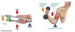
Third-class lever: Force between fulcrum and load (e.g., lifting a dumbbell with the forearm).
Mechanical Advantage and Disadvantage
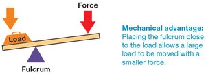
Mechanical Advantage: When the fulcrum is farther from the force, a small force can move a large load over a short distance.
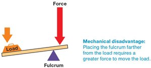
Mechanical Disadvantage: When the fulcrum is close to the force, a greater force is needed to move the load, but the load moves faster and over a greater distance.
Muscles of the Head, Neck, and Vertebral Column
Facial Muscles
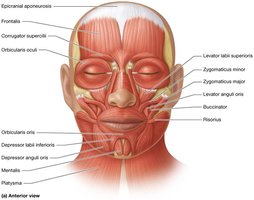
Muscles of facial expression allow for a wide range of movements, including smiling, frowning, and blinking. These muscles are unique because they insert into the skin rather than bone.
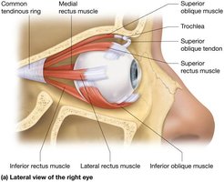
Extrinsic Eye Muscles
These muscles control the movement of the eyeball and are responsible for precise eye positioning.
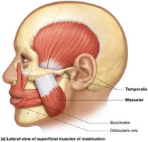
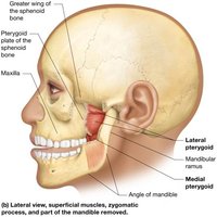
Muscles of Mastication
These muscles are responsible for chewing and moving the jaw.
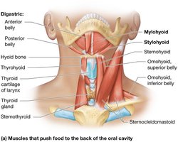
Muscles of Swallowing
These muscles coordinate the movement of food from the mouth to the esophagus.
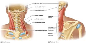
Muscles That Move the Head and Neck
These muscles allow for flexion, extension, rotation, and lateral movement of the head and neck.
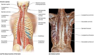
Muscles of the Vertebral Column
These muscles support the spine and allow for its movement and stabilization.
Muscles of the Trunk and Pelvic Floor
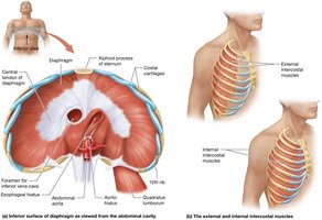
Muscles of Respiration
The diaphragm and intercostal muscles are essential for breathing by changing the volume of the thoracic cavity.
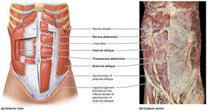
Abdominal Muscles
These muscles support the abdominal organs, assist in breathing, and help with trunk movement and stability.
Muscles of the Pelvic Floor and Perineum
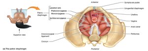
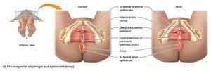
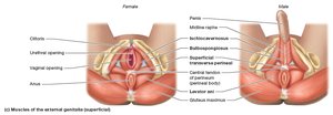
The pelvic floor muscles support pelvic organs and control the openings of the urethra, vagina, and anus.
Muscles of the Pectoral Girdle and Upper Limbs
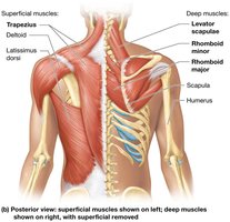
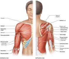
Muscles That Move the Scapula and Arm
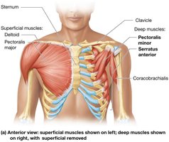
These muscles stabilize and move the shoulder blade and upper arm, allowing for a wide range of arm movements.
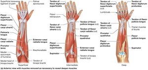
Muscles of the Forearm and Hand
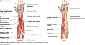
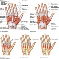
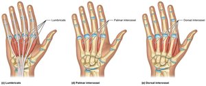
These muscles control movements of the wrist, hand, and fingers, enabling fine motor skills.
Muscles of the Hip and Lower Limb
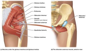
Muscles That Move the Thigh and Knee
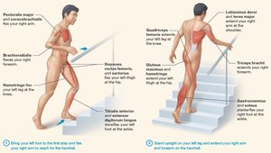
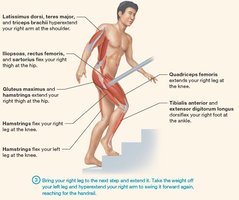
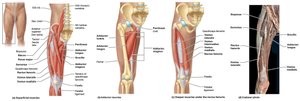
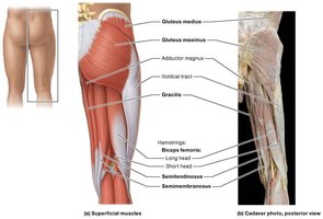
These muscles are responsible for movements such as walking, running, and jumping by acting on the hip and knee joints.
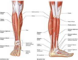
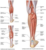
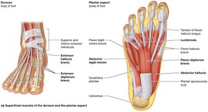
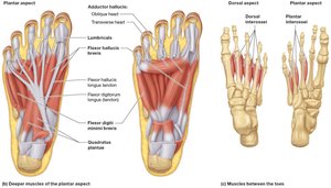
Muscles That Move the Ankle and Foot
These muscles allow for dorsiflexion, plantarflexion, inversion, and eversion of the foot, as well as toe movements.
Integration of Muscle Function
Coordinated Movement
Complex body movements require the coordinated action of multiple muscle groups, each contributing to stability, force, and precision.