Back
BackThe Muscular System: Structure, Function, and Clinical Applications
Study Guide - Smart Notes
 Tailored notes based on your materials, expanded with key definitions, examples, and context.
The Muscular System
Introduction
The muscular system is essential for movement, posture, and various bodily functions. This chapter focuses on the structure, organization, and function of skeletal muscles, as well as clinical considerations related to muscle injuries and disorders.
Structure of Skeletal Muscles
Gross Anatomy of a Skeletal Muscle
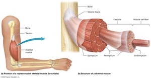
Skeletal Muscle Fibers: Long, thin cells surrounded by a thin layer of extracellular matrix called the endomysium.
Fascicle: A bundle of 10–100 muscle fibers, surrounded by perimysium.
Epimysium: Connective tissue that surrounds all fascicles in a muscle; continuous with the fascia, the most superficial connective tissue sheath.
Tendons: Attach muscle to bone or other structures to facilitate movement.
Voluntary Control: Skeletal muscle contraction is under conscious control and requires nervous system stimulation.
Muscle Knots (Myofascial Trigger Points)
Definition and Clinical Relevance
Myofascial Trigger Point: A localized, irritable spot within the fascia surrounding a muscle, often inflamed and painful.
Causes: Repetitive exercise, trauma, stress, disease, or trapped nerves can lead to trigger points.
Treatment: Includes massage, anti-inflammatory medications, muscle relaxants, and passive stretching.
Fascicle Patterns and Muscle Shapes
Classification of Muscle Shapes
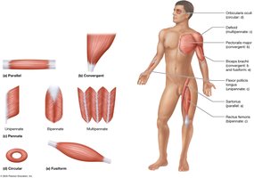
Parallel: Evenly spaced fascicles attaching to a tendon of similar width.
Convergent: Broad at one end, tapering to a single tendon.
Circular (Sphincters): Encircle openings, constricting them when contracted.
Fusiform: Thick in the middle, tapered at the ends.
Pennate: Fascicles attach to the tendon at an angle, resembling a feather.
Unipennate: Fascicles on one side of the tendon.
Bipennate: Fascicles on both sides of the tendon.
Multipennate: Multiple tendons with fascicles arranged like several feathers.
Naming Muscles
Principles of Muscle Nomenclature
Size: Terms like major, minor, longus, brevis, and vastus.
Location: Directional (superior, inferior, medial, lateral) and regional anatomical terms.
Attachment: Named for structures to which they attach.
Function: Flexors, extensors, levators, adductors, and abductors.
Depth: Terms like profundus (deep) and superficialis (superficial).
Functional Groups of Muscles
Roles in Movement
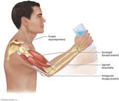
Agonist (Prime Mover): Provides most of the force for a movement.
Antagonist: Opposes and slows the action of the agonist; important for controlled movements.
Synergist: Assists the agonist, guiding and smoothing the movement.
Fixator: Stabilizes a bone, making movement more efficient and reducing injury risk.
Muscle Origin and Insertion
Attachment Sites
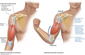
Origin (O): The more fixed attachment point of a muscle.
Insertion (I): The attachment point that moves during contraction.
Example: The biceps brachii originates on the scapula (stationary) and inserts on the radius (moves).
Lever Systems Used in Body Movements
Components and Types of Levers
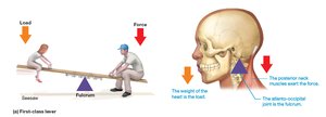
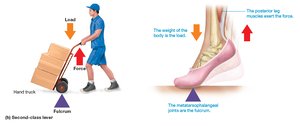
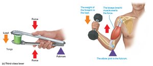
Lever: The bone that moves.
Load: The object or body part being moved.
Force: Muscle tension applied to the lever.
Fulcrum: The joint around which the lever moves.
There are three classes of levers, distinguished by the relative positions of the fulcrum, load, and force:
First-class lever: Fulcrum is between the load and the applied force (e.g., seesaw, nodding head).
Second-class lever: Load is between the fulcrum and the applied force (e.g., wheelbarrow, standing on tiptoe).
Third-class lever: Force is between the fulcrum and the load (e.g., tweezers, biceps curl).
Muscle Strains
Definition, Symptoms, and Treatment
Muscle Strain: Occurs when a muscle is overstretched or subjected to excessive force, resulting in tearing of muscle fibers.
Symptoms: Pain, limited movement, swelling, and bruising.
Treatment: The PRICE method (Protect, Rest, Ice, Compression, Elevation), anti-inflammatory medications, physical therapy, or surgery for severe cases.
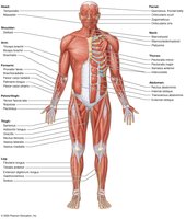
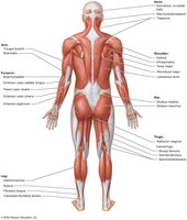
Superficial Muscles of the Body
Anterior and Posterior Views
Superficial muscles are those closest to the skin and are responsible for the majority of visible muscle contours.
Understanding their locations is essential for clinical assessment and anatomical study.
Muscles of Facial Expression
Eyes and Mouth
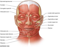
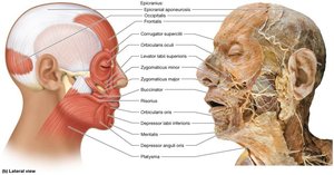
Muscles of facial expression insert into the skin and connective tissue of the face, allowing for subtle changes in appearance.
Epicranial aponeurosis: Connects the frontalis and occipitalis muscles, raising the eyebrows and forehead skin.
Orbicularis oculi: Closes the eyelids.
Zygomaticus major/minor, levator labii superioris, risorius, orbicularis oris, depressor anguli oris, depressor labii inferioris, mentalis, buccinator, platysma: Control movements of the mouth, lips, and chin, and convey emotions.
Extrinsic Eye and Orbit Muscles
Rectus and Oblique Muscles
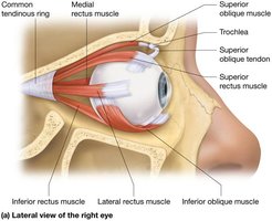
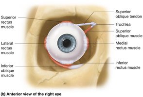
Each eye has six extrinsic muscles for precise movement.
Rectus muscles: Superior, inferior, medial, and lateral rectus move the eye up, down, and side-to-side.
Oblique muscles: Superior and inferior oblique rotate the eye and assist in complex movements.
Muscles of Chewing (Mastication)
Major Muscles and Actions
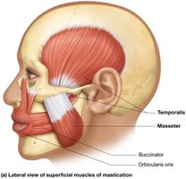
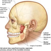
Masseter and temporalis: Elevate the mandible for biting and chewing.
Medial and lateral pterygoid: Assist in elevation, protraction, and side-to-side movement of the mandible.
Muscles of Swallowing
Oral Cavity, Pharynx, and Esophagus
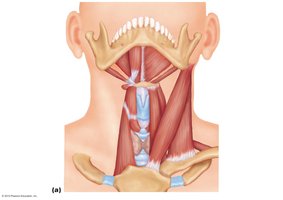
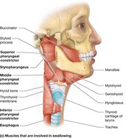
Tongue muscles: Genioglossus, hyoglossus, styloglossus manipulate food and initiate swallowing.
Pharyngeal muscles: Sternohyoid, sternothyroid, omohyoid, thyrohyoid, pharyngeal constrictors, digastric, and stylohyoid coordinate swallowing and movement of food into the esophagus.
Muscles that Move the Head and Neck
Major Muscles
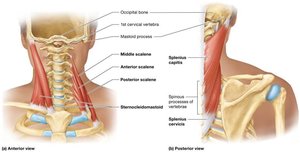
Sternocleidomastoid, scalenes, trapezius, splenius capitis, splenius cervicis, semispinalis capitis: Responsible for flexion, extension, rotation, and lateral bending of the head and neck.
Muscles of the Vertebral Column
Posture and Locomotion
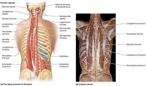
Erector spinae group: Spinalis (medial), longissimus (intermediate), iliocostalis (lateral) – extend and laterally bend the spine.
Quadratus lumborum: Extends and bends the vertebral column laterally.
Muscles of the Trunk
Respiration and Abdominal Wall
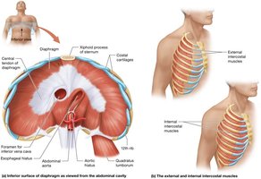
Muscles of respiration: Diaphragm, internal intercostals, external intercostals.
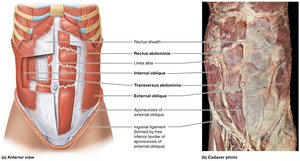
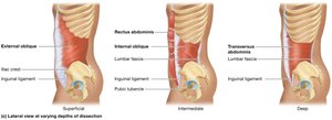
Abdominal muscles: Rectus abdominis, external obliques, internal obliques, transversus abdominis; linea alba separates left and right rectus muscles.
Muscles of the Pelvic Floor and Perineum
Pelvic Diaphragm, Urogenital Diaphragm, and Perineum
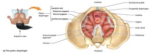
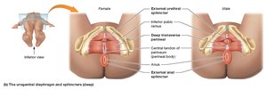
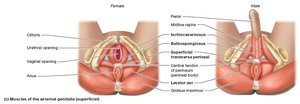
Pelvic diaphragm: Levator ani group (pubococcygeus, iliococcygeus), coccygeus – support pelvic organs.
Urogenital diaphragm: External urethral sphincter, deep and superficial transverse perineal muscles – support pelvic region and control urination.
Perineum: Bulbospongiosus and ischiocavernosus – support external genitalia.
Urinary Incontinence and Kegel Exercises
Clinical Application
Urinary incontinence: Often due to weak pelvic diaphragm muscles, especially after childbirth or with aging.
Kegel exercises: Strengthen the levator ani muscle to treat incontinence by repeated contraction and relaxation.

Muscles that Move the Scapula, Arm, and Forearm
Major Muscle Groups
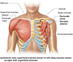
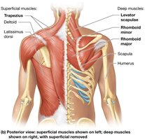
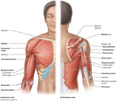
Muscles such as the trapezius, deltoid, pectoralis major, and rotator cuff muscles are responsible for movements of the scapula, arm, and forearm.
Rotator Cuff Injuries
Clinical Considerations
Rotator cuff muscles: Stabilize the shoulder joint during overhead movements.
Injury risk: Repetitive overhead activity, anatomical variations (e.g., hooked acromion), or bone spurs can cause tears.
Symptoms: Pain, weakness, decreased range of motion.
Treatment: Rest, anti-inflammatory medications, physical therapy, or surgery.

Muscles that Move the Hand, Thigh, Leg, Foot, and Toes
Overview of Major Muscle Groups
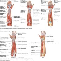
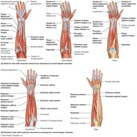
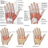
Muscles of the forearm and hand control fine movements and grip strength.
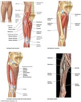
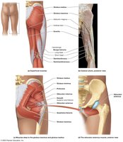
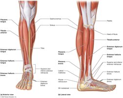
Muscles of the thigh and leg are responsible for locomotion, balance, and posture.
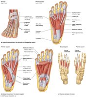
Muscles of the foot and toes provide stability and movement during walking and running.
Calcaneal Tendon Injuries
Clinical Considerations
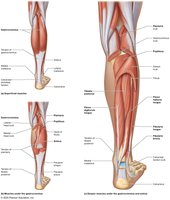
Gastrocnemius and soleus: Form a second-class lever, generating significant force for plantarflexion.
Injury risk: Common in sports; includes tendonitis, partial, or complete tears.
Diagnosis: Simmonds Test (absence of plantarflexion when tendon is ruptured).
Treatment: May include immobilization, physical therapy, or surgery.

Muscles that Move the Toes
Overview
Muscles of the foot are responsible for flexion, extension, abduction, and adduction of the toes, contributing to balance and locomotion.