 Back
BackThe Nervous System and Nervous Tissue: Structure, Function, and Integration
Study Guide - Smart Notes
 Tailored notes based on your materials, expanded with key definitions, examples, and context.
The Nervous System: Overview and Divisions
Introduction to the Nervous System
The nervous system is a complex network responsible for receiving, processing, and transmitting information throughout the body. It consists of the brain, spinal cord, sensory receptors, and nerves. The nervous system is essential for regulating bodily functions, responding to stimuli, and maintaining homeostasis.
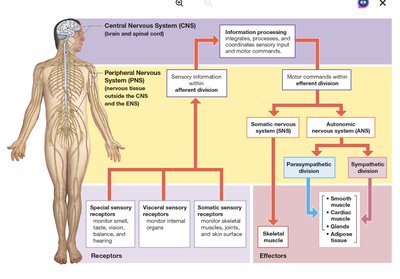
Central Nervous System (CNS): Composed of the brain and spinal cord; integrates and processes sensory data and motor commands.
Peripheral Nervous System (PNS): Includes all neural tissue outside the CNS; delivers sensory information to the CNS and carries motor commands to peripheral tissues.
Additional info: The CNS is also the seat of higher functions such as intelligence, memory, and emotion.
Anatomical and Functional Divisions
Anatomical Divisions: CNS (brain and spinal cord) and PNS (nerves and ganglia).
Functional Divisions of the PNS:
Afferent Division: Brings sensory information from receptors to the CNS.
Efferent Division: Carries motor commands from the CNS to effectors (muscles, glands, adipose tissue).
Somatic Nervous System (SNS): Controls voluntary and involuntary skeletal muscle contractions.
Autonomic Nervous System (ANS): Regulates involuntary functions (smooth muscle, cardiac muscle, glands, adipose tissue) and includes sympathetic and parasympathetic divisions.
Enteric Nervous System (ENS): Network of neurons in the digestive tract, capable of local reflexes independent of the CNS.
Neurons: Structure and Classification
Structure of a Typical Neuron
Neurons are the basic functional units of the nervous system, specialized for intercellular communication. They are generally long-lived, have a high metabolic rate, and possess an excitable plasma membrane.
Cell Body (Soma): Contains the nucleus, nucleolus, and organelles such as mitochondria, ribosomes, and rough endoplasmic reticulum (RER). Nissl bodies (clusters of RER and ribosomes) give gray matter its color.
Dendrites: Highly branched extensions that receive information from other neurons.
Axon: Long process that propagates action potentials; contains axoplasm and is surrounded by the axolemma. The axon hillock is the origin of the axon.
Telodendria: Terminal branches ending in axon terminals (synaptic terminals) that communicate with other cells.
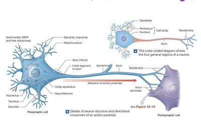
Additional info: Axonal transport moves materials between the cell body and axon terminals via molecular motors (kinesin for anterograde, dynein for retrograde transport).
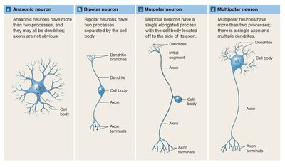
Structural Classification of Neurons
Anaxonic Neurons: Small, with many dendrites but no obvious axon; found in the brain and special sense organs.
Bipolar Neurons: Have one dendrite and one axon with the cell body in between; rare, found in special sense organs.
Unipolar Neurons: Dendrites and axon are continuous; cell body off to one side; most sensory neurons in the PNS are unipolar.
Multipolar Neurons: Two or more dendrites and a single axon; most common in the CNS and all motor neurons controlling skeletal muscles.
Functional Classification of Neurons
Sensory Neurons (Afferent): Deliver information from sensory receptors to the CNS; cell bodies located in sensory ganglia.
Motor Neurons (Efferent): Carry instructions from the CNS to effectors; include somatic motor neurons (skeletal muscle) and visceral motor neurons (smooth/cardiac muscle, glands, adipose tissue).
Interneurons: Located between sensory and motor neurons; responsible for integration, coordination, and higher functions such as memory and learning.
Additional info: Sensory receptors are classified as interoceptors (internal environment), exteroceptors (external environment), and proprioceptors (position and movement).
Neuroglia: Support Cells of the Nervous System
Types and Functions of Neuroglia
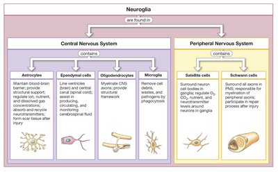
Neuroglia are supporting cells that separate and protect neurons, provide structural support, act as phagocytes, and regulate the interstitial environment. They outnumber neurons and are essential for nervous system function.
Central Nervous System | Peripheral Nervous System |
|---|---|
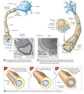
Astrocytes: Maintain blood-brain barrier, provide structural support, regulate ion/nutrient/gas concentrations, absorb/recycle neurotransmitters, form scar tissue after injury. Ependymal Cells: Line ventricles and central canal, produce/monitor/circulate cerebrospinal fluid. Oligodendrocytes: Myelinate CNS axons, provide structural framework. Microglia: Remove cell debris, wastes, and pathogens by phagocytosis. | Satellite Cells: Surround neuron cell bodies in ganglia, regulate interstitial fluid. Schwann Cells: Surround all axons in PNS, responsible for myelination and repair after injury. |
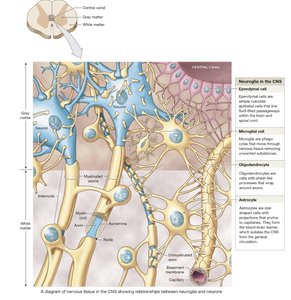
Myelination and Repair
Myelin Sheath: Formed by oligodendrocytes in the CNS and Schwann cells in the PNS; increases speed of action potential conduction and insulates axons.
Nodes of Ranvier: Gaps between myelinated segments where action potentials are regenerated.
Wallerian Degeneration: In the PNS, Schwann cells assist in the repair of damaged axons by forming a regeneration tube.

Additional info: Demyelinating diseases such as multiple sclerosis and diphtheria disrupt normal nerve function.
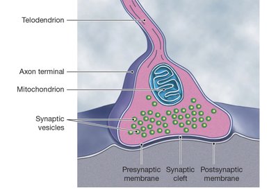
Synapses and Neurotransmission
Structure and Function of Synapses
A synapse is a specialized junction where a neuron communicates with another cell. Synapses can be electrical (direct ion flow via gap junctions) or chemical (neurotransmitter release across a synaptic cleft).
Presynaptic Neuron: Releases neurotransmitters from synaptic vesicles in the axon terminal.
Postsynaptic Cell: Receives the signal via receptors on its membrane.
Synaptic Cleft: The small gap between the presynaptic and postsynaptic membranes.
Chemical Synaptic Transmission (Cholinergic Synapse Example)
An action potential arrives at the presynaptic axon terminal and depolarizes the membrane.
Voltage-gated calcium channels open, and Ca2+ enters the terminal, triggering exocytosis of acetylcholine (ACh) from synaptic vesicles.
ACh diffuses across the synaptic cleft and binds to receptors on the postsynaptic membrane, causing depolarization (EPSP).
ACh is broken down by acetylcholinesterase (AChE), terminating the signal.
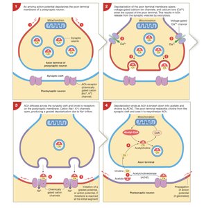
Additional info: Synaptic delay and synaptic fatigue can affect the efficiency of neurotransmission.
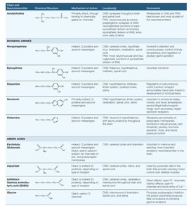
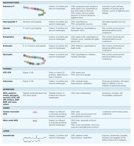
Neurotransmitters and Neuromodulators
Major Classes and Effects
Neurotransmitters are chemicals that transmit signals across synapses. Their effects depend on the receptors they bind to, not just their chemical nature. Neuromodulators alter the response of neurons to neurotransmitters.
Class | Examples | Primary Effects |
|---|---|---|
Biogenic Amines | Norepinephrine, Dopamine, Serotonin, Histamine | Excitatory or inhibitory, depending on receptor type |
Amino Acids | Glutamate, Aspartate, GABA, Glycine | Glutamate/aspartate: excitatory; GABA/glycine: inhibitory |
Neuropeptides | Substance P, Opioids (endorphins, enkephalins, dynorphins) | Pain modulation, neuromodulation |
Dissolved Gases | Nitric oxide (NO), Carbon monoxide (CO) | Indirect effects via intracellular enzymes |
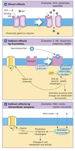
Mechanisms of Action
Direct Effects: Open or close chemically gated ion channels (ionotropic effects), e.g., ACh, glutamate.
Indirect Effects via G Proteins: Activate second messengers (e.g., cAMP) through G protein-coupled receptors (metabotropic effects), e.g., norepinephrine, dopamine.
Indirect Effects via Intracellular Enzymes: Lipid-soluble gases diffuse into cells and activate enzymes, e.g., NO, CO.
Neural Integration and Information Processing
Postsynaptic Potentials and Summation
Neurons integrate excitatory and inhibitory inputs to determine whether an action potential will be generated. This integration occurs primarily at the axon hillock.
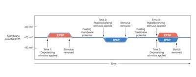
Excitatory Postsynaptic Potential (EPSP): Graded depolarization that increases the likelihood of an action potential.
Inhibitory Postsynaptic Potential (IPSP): Graded hyperpolarization that decreases the likelihood of an action potential.
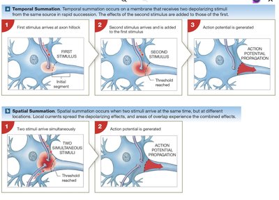
Summation: The process by which multiple EPSPs and IPSPs combine to influence the membrane potential.
Temporal Summation: Rapid, repeated stimuli at a single synapse.
Spatial Summation: Simultaneous stimuli at multiple synapses.
Presynaptic Regulation and Action Potential Frequency
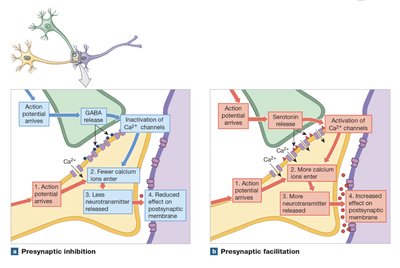
Presynaptic Inhibition: Reduces neurotransmitter release (e.g., GABA-mediated inhibition of Ca2+ channels).
Presynaptic Facilitation: Increases neurotransmitter release (e.g., serotonin prolongs Ca2+ channel opening).
Action Potential Frequency: The rate of action potential generation encodes the intensity of a stimulus; higher depolarization leads to higher frequency.
Additional info: The absolute refractory period limits the maximum frequency of action potentials.
