 Back
BackThe Nervous System: Structure and Anatomy of the Brain, Spinal Cord, and Special Senses
Study Guide - Smart Notes
 Tailored notes based on your materials, expanded with key definitions, examples, and context.
The Nervous System
Overview of the Nervous System
The nervous system is a complex network responsible for coordinating the body's activities by transmitting signals to and from different parts of the body. It is divided into the central nervous system (CNS), which includes the brain and spinal cord, and the peripheral nervous system (PNS), which consists of nerves and ganglia outside the CNS.
Central Nervous System (CNS): Composed of the brain and spinal cord, responsible for processing and integrating information.
Peripheral Nervous System (PNS): Includes all neural tissue outside the CNS, connecting the CNS to limbs and organs.
Major Functions: Sensory input, integration, motor output, and homeostasis regulation.
Gross Anatomy of the Brain
Major Regions of the Brain
The brain is divided into several major regions, each with specialized functions. Understanding the anatomical organization is essential for studying neural function and clinical neuroanatomy.
Cerebrum: The largest part of the brain, responsible for higher brain functions such as thought, action, and sensory processing.
Cerebellum: Located under the cerebrum, it coordinates voluntary movements, balance, and posture.
Diencephalon: Contains structures such as the thalamus and hypothalamus, involved in sensory relay and homeostatic regulation.
Brainstem: Includes the midbrain, pons, and medulla oblongata; controls basic life functions such as breathing, heart rate, and digestion.
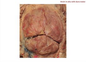
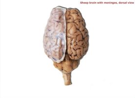
Meninges and Brain Protection
The brain is protected by three connective tissue membranes called meninges: dura mater, arachnoid mater, and pia mater. The dura mater is the tough outermost layer, providing a durable protective covering.
Dura Mater: Outermost, tough, and fibrous layer.
Arachnoid Mater: Middle, web-like layer.
Pia Mater: Innermost, delicate layer adhering closely to the brain surface.
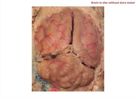
Surface Anatomy of the Brain
The brain's surface is marked by gyri (ridges) and sulci (grooves), increasing surface area for neural processing. The longitudinal fissure divides the brain into right and left cerebral hemispheres, while the transverse fissure separates the cerebrum from the cerebellum.
Gyri: Elevated ridges on the brain surface.
Sulci: Shallow grooves between gyri.
Fissures: Deep grooves, such as the longitudinal and transverse fissures.
Internal Anatomy of the Brain
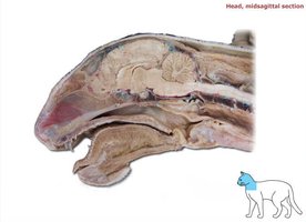
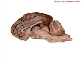
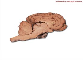
Midsagittal Section of the Brain
A midsagittal section reveals the internal organization of the brain, including the corpus callosum, ventricles, and brainstem structures. This view is essential for understanding the relationships between major brain regions.
Corpus Callosum: Thick band of nerve fibers connecting the two cerebral hemispheres.
Ventricles: Cavities within the brain filled with cerebrospinal fluid (CSF), including the lateral, third, and fourth ventricles.
Arbor Vitae: Tree-like white matter structure in the cerebellum.
Thalamus and Hypothalamus: Key diencephalic structures involved in sensory relay and autonomic control.

External Views of the Brain
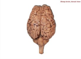
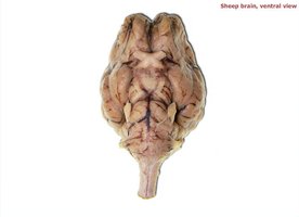
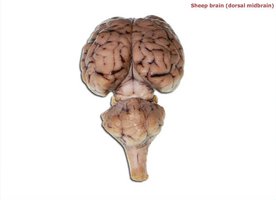
Dorsal and Ventral Views
Examining the brain from different perspectives helps identify major external features and the arrangement of cranial nerves and fissures.
Dorsal View: Shows the two cerebral hemispheres, longitudinal fissure, and cerebellum.
Ventral View: Reveals the cranial nerves, optic chiasm, and brainstem structures.
Peripheral Nervous System: Nerve Plexuses
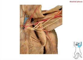
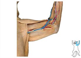
Brachial Plexus
The brachial plexus is a network of nerves that innervates the muscles and skin of the shoulder, arm, and hand. It is formed by the ventral rami of spinal nerves C5-T1.
Major Nerves: Median, ulnar, radial, and musculocutaneous nerves.
Function: Motor and sensory innervation to the upper limb.
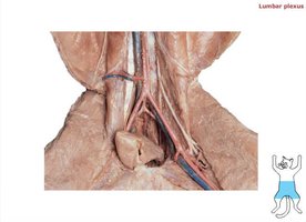
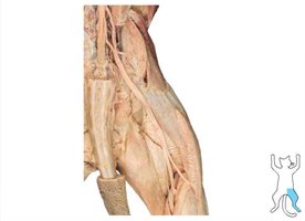
Lumbar and Sacral Plexuses
The lumbar and sacral plexuses supply the lower limb. The lumbar plexus arises from L1-L4, while the sacral plexus arises from L4-S4.
Lumbar Plexus: Includes the femoral, obturator, and lateral femoral cutaneous nerves.
Sacral Plexus: Includes the sciatic, tibial, and common fibular nerves.
Function: Motor and sensory innervation to the lower limb and pelvic region.
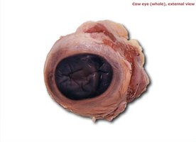
Special Senses: The Eye
Gross Anatomy of the Eye
The eye is a specialized organ for vision, composed of three layers: the fibrous tunic (sclera and cornea), vascular tunic (choroid, ciliary body, iris), and nervous tunic (retina). The lens focuses light onto the retina, and the optic nerve transmits visual information to the brain.
Sclera: Tough, white outer layer providing protection.
Cornea: Transparent anterior portion allowing light entry.
Lens: Biconvex structure that focuses light.
Retina: Light-sensitive layer containing photoreceptors.
Optic Nerve: Transmits visual signals to the brain.
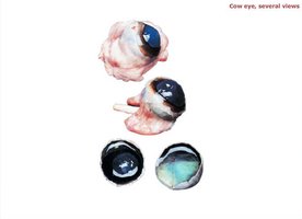
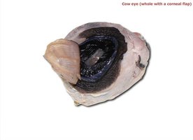
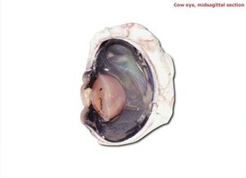
Internal Structures of the Eye
A midsagittal section of the eye reveals the arrangement of internal structures, including the lens, iris, pupil, choroid, and vitreous humor. These components work together to regulate light entry and focus images on the retina.
Iris: Colored part of the eye, controls pupil size.
Pupil: Opening in the iris through which light enters.
Choroid: Vascular layer providing nourishment to the retina.
Vitreous Humor: Gel-like substance filling the posterior cavity, maintaining eye shape.
Summary Table: Major Brain Regions and Functions
Region | Main Structures | Primary Functions |
|---|---|---|
Cerebrum | Cerebral cortex, corpus callosum | Conscious thought, sensory processing, voluntary movement |
Cerebellum | Arbor vitae, hemispheres | Coordination, balance, posture |
Diencephalon | Thalamus, hypothalamus, pineal gland | Sensory relay, autonomic control, hormone regulation |
Brainstem | Midbrain, pons, medulla oblongata | Basic life functions (breathing, heart rate, digestion) |
