 Back
BackThe Nervous System: Structure and Function
Study Guide - Smart Notes
 Tailored notes based on your materials, expanded with key definitions, examples, and context.
The Nervous System
Overview of the Nervous System
The nervous system is a complex network responsible for coordinating the body's activities by transmitting signals to and from different parts of the body. It is divided into the central nervous system (CNS) and the peripheral nervous system (PNS).
Central Nervous System (CNS): Consists of the brain and spinal cord; processes information and determines responses.
Peripheral Nervous System (PNS): Composed of nerves and ganglia outside the CNS; transmits signals between the CNS and the rest of the body.
Neurons and Neuroglial Cells
Structure and Types of Neurons
Neurons are the primary functional units of the nervous system, specialized for the transmission of electrical impulses.
Cell Body (Soma): Contains the nucleus and organelles; responsible for metabolic activities.
Dendrites: Receive incoming signals from other neurons.
Axon: Conducts electrical impulses away from the cell body toward other neurons or effectors.
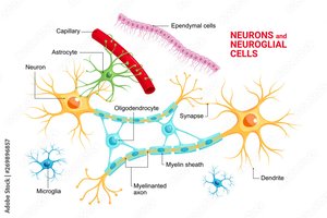
Neuroglial Cells
Neuroglia (glial cells) support, protect, and nourish neurons. Major types include:
Astrocytes: Maintain the blood-brain barrier and provide structural support.
Oligodendrocytes: Form myelin sheaths in the CNS.
Schwann Cells: Form myelin sheaths in the PNS.
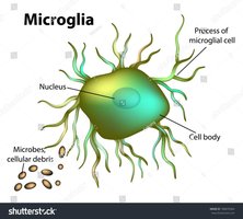
Microglia: Act as immune cells, removing debris and pathogens.
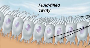
Ependymal Cells: Line fluid-filled cavities in the CNS and help circulate cerebrospinal fluid.
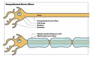
Myelinated vs. Unmyelinated Nerve Fibers
Structure and Function
Nerve fibers can be myelinated or unmyelinated, affecting the speed of nerve impulse conduction.
Myelinated Fibers: Axons covered with a myelin sheath, which increases the speed of impulse transmission.
Unmyelinated Fibers: Axons without a myelin sheath; impulses travel more slowly.
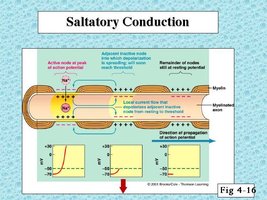
Saltatory Conduction
In myelinated axons, action potentials jump from one node of Ranvier to the next, greatly increasing conduction speed.
Saltatory Conduction: The process by which action potentials leap between nodes of Ranvier in myelinated fibers.
Continuous Conduction: Occurs in unmyelinated fibers, where the action potential travels along every part of the membrane.
Information Flow in Neurons
Direction of Signal Transmission
Neurons transmit information in a unidirectional manner:
Dendrites: Receive signals.
Cell Body: Integrates incoming signals.
Axon: Conducts the action potential to the axon terminals.

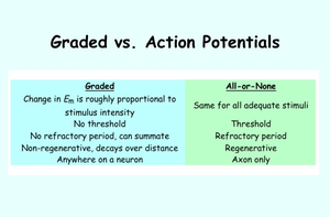
Electrical Signals: Graded and Action Potentials
Graded Potentials
Graded potentials are changes in membrane potential that vary in size and decay with distance.
Occur in dendrites and cell bodies.
Can summate to trigger an action potential if threshold is reached.
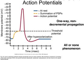
Action Potentials
Action potentials are rapid, all-or-none electrical impulses that travel along axons.
Triggered when membrane potential reaches threshold.
Exhibit a refractory period and do not decay over distance.
Graded Potentials | Action Potentials |
|---|---|
Proportional to stimulus intensity | All-or-none for adequate stimuli |
No threshold | Threshold required |
No refractory period | Refractory period present |
Decay over distance | Regenerative, do not decay |
Anywhere on neuron | Axon only |
Action Potential Generation and Propagation
The action potential is generated at the axon hillock and propagates along the axon.
Depolarization: Na+ influx causes the membrane potential to become more positive.
Repolarization: K+ efflux restores the negative membrane potential.
Hyperpolarization: Membrane potential becomes more negative than resting potential before stabilizing.
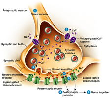
Synaptic Transmission
Chemical Synapses
At chemical synapses, neurotransmitters are released from the presynaptic neuron and bind to receptors on the postsynaptic cell, initiating a new electrical signal.
Presynaptic Terminal: Releases neurotransmitter in response to an action potential.
Synaptic Cleft: The gap between neurons where neurotransmitters diffuse.
Postsynaptic Membrane: Contains receptors that bind neurotransmitters and generate a response.
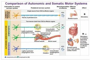
Autonomic vs. Somatic Motor Systems
Comparison of Motor Pathways
The somatic and autonomic nervous systems differ in structure, neurotransmitters, and effectors.
Somatic Motor System: Controls voluntary movements via skeletal muscle; uses a single neuron pathway and acetylcholine (ACh) as the neurotransmitter.
Autonomic Motor System: Controls involuntary functions (e.g., heart, glands, smooth muscle); uses a two-neuron chain and various neurotransmitters (ACh, norepinephrine).
System | Neural Pathway | Neurotransmitter | Effector | Effect |
|---|---|---|---|---|
Somatic | Single, heavily myelinated axon | ACh | Skeletal muscle | Stimulatory |
Autonomic (Sympathetic) | Two-neuron chain | ACh, NE | Cardiac/smooth muscle, glands | Stimulatory or inhibitory |
Autonomic (Parasympathetic) | Two-neuron chain | ACh | Cardiac/smooth muscle, glands | Stimulatory or inhibitory |
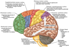
Functional Areas of the Brain
Cerebral Cortex Regions
The brain contains specialized regions responsible for different functions.
Primary Motor Cortex: Controls voluntary movements.
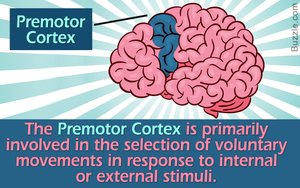
Premotor Cortex: Involved in planning and selecting movements in response to stimuli.
Prefrontal Cortex: Responsible for complex behaviors, decision-making, and personality.
Somatosensory Cortex: Processes sensory input from the body.
Cerebellum: Coordinates movement and balance.
Summary Table: Key Concepts
Concept | Description |
|---|---|
Neuron | Basic functional unit of the nervous system |
Neuroglia | Support cells for neurons |
Myelin | Insulating sheath that increases conduction speed |
Action Potential | All-or-none electrical impulse along axon |
Synapse | Junction between two neurons |
Somatic System | Controls voluntary muscles |
Autonomic System | Controls involuntary functions |
Additional info: This guide integrates foundational concepts in nervous system anatomy and physiology, including neuron structure, neuroglia, electrical signaling, synaptic transmission, and functional brain regions, as relevant to introductory college-level ANP courses.
